Tuesday Poster Session
Category: Biliary/Pancreas
P3515 - Intrapapillary Mucinous Neoplasm Complicated by Spontaneous Pancreaticogastric Fistula
Tuesday, October 29, 2024
10:30 AM - 4:00 PM ET
Location: Exhibit Hall E

Has Audio
- MK
Muhammad Zarrar Khan, MD
Henry Ford Health
Detroit, MI
Presenting Author(s)
Krishna Vemulapalli, MD1, Muhammad Z. Khan, MD2, Palak Patel-Rodrigues, MD2, Tariq Hammad, MD1, Andrew Watson, MD2
1Henry Ford Hospital, Detroit, MI; 2Henry Ford Health, Detroit, MI
Introduction: Intraductal Papillary Mucinous Neoplasms (IPMNs) consist of a range of presentations varying in malignant potential. Mainly characterized by papillary growth and significant mucin secretion, IPMNs can present with various complications including obstructive jaundice or cholangitis. Rarely, IPMN can fistulize into the adjacent organs. Here we present the case of a patient found to have benign IPMN complicated by 20mm pancreatico-gastric fistula.
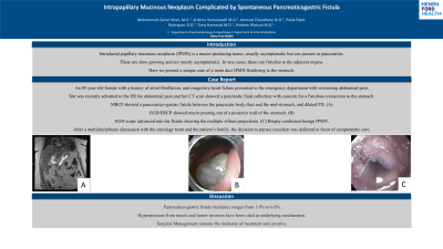
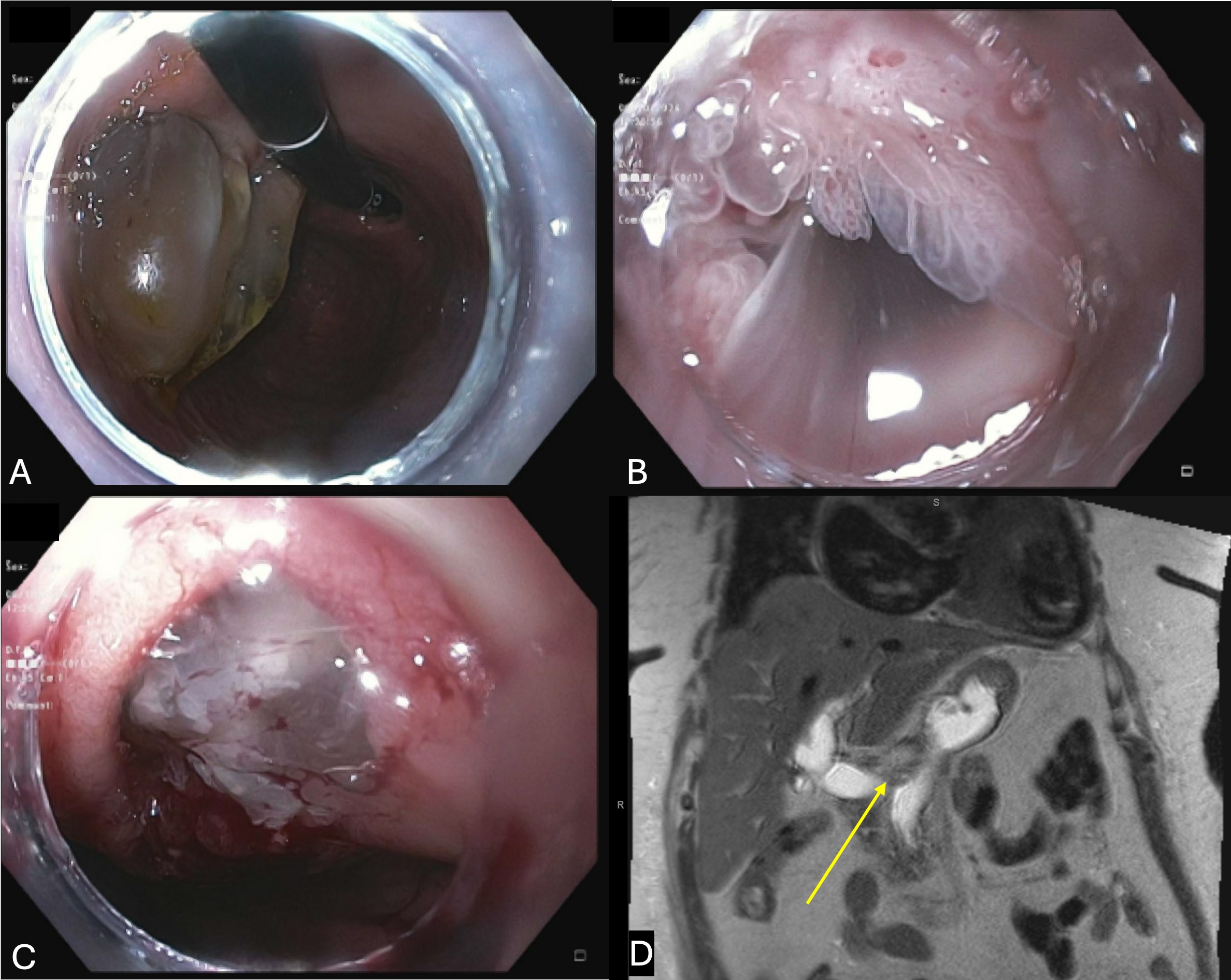
Case Description/Methods: An 85-year-old female with history of heart failure presented with several week history of progressive epigastric pain accompanied by poor appetite and nausea. She remained hemodynamically stable with lab work showing unremarkable liver enzymes, blood counts, and lipase. CT Abdomen Pelvis showed dilated pancreatic duct with communication to the stomach. Magnetic resonance cholangiopancreatography (MRCP) showed moderate intrahepatic and severe extrahepatic biliary ductal dilation with the main pancreatic duct dilated up to 20mm. A 20mm fistula between the pancreatic body duct to posterior wall of the stomach was noted. The patient underwent EGD/ERCP which demonstrated a 20mm fistula with copious amounts of mucin pouring out of the lesser curvature opening. The main pancreatic duct was visualized with features of main duct IPMN. Biopsy demonstrated features consistent with low-grade IPMN. Sphincterotomy and balloon extraction of choledocholithiasis was performed. A fully covered metal stent was placed into the common bile duct to maintain biliary drainage. Multidisciplinary discussion was held with the patient’s family, gastroenterology team, and oncology team and decision to pursue resection was deferred in favor of symptomatic care. The patient recovered well and was discharged to follow-up for outpatient ERCP for further stent management.
Discussion: IPMN resulting in fistulation into the stomach is an exceedingly rare presentation that can mimic many other pathologies of abdominal pain. Our patient’s presentation with non-specific symptoms of epigastric pain, nausea, and early satiety requires a high degree of suspicion for biliary pathology. The identification of such a diagnosis requires complex decision-making involving multidisciplinary insight as well as shared decision-making regarding the potential for malignancy and surgical versus conservative management.

Disclosures:
Krishna Vemulapalli, MD1, Muhammad Z. Khan, MD2, Palak Patel-Rodrigues, MD2, Tariq Hammad, MD1, Andrew Watson, MD2. P3515 - Intrapapillary Mucinous Neoplasm Complicated by Spontaneous Pancreaticogastric Fistula, ACG 2024 Annual Scientific Meeting Abstracts. Philadelphia, PA: American College of Gastroenterology.
1Henry Ford Hospital, Detroit, MI; 2Henry Ford Health, Detroit, MI
Introduction: Intraductal Papillary Mucinous Neoplasms (IPMNs) consist of a range of presentations varying in malignant potential. Mainly characterized by papillary growth and significant mucin secretion, IPMNs can present with various complications including obstructive jaundice or cholangitis. Rarely, IPMN can fistulize into the adjacent organs. Here we present the case of a patient found to have benign IPMN complicated by 20mm pancreatico-gastric fistula.
Case Description/Methods: An 85-year-old female with history of heart failure presented with several week history of progressive epigastric pain accompanied by poor appetite and nausea. She remained hemodynamically stable with lab work showing unremarkable liver enzymes, blood counts, and lipase. CT Abdomen Pelvis showed dilated pancreatic duct with communication to the stomach. Magnetic resonance cholangiopancreatography (MRCP) showed moderate intrahepatic and severe extrahepatic biliary ductal dilation with the main pancreatic duct dilated up to 20mm. A 20mm fistula between the pancreatic body duct to posterior wall of the stomach was noted. The patient underwent EGD/ERCP which demonstrated a 20mm fistula with copious amounts of mucin pouring out of the lesser curvature opening. The main pancreatic duct was visualized with features of main duct IPMN. Biopsy demonstrated features consistent with low-grade IPMN. Sphincterotomy and balloon extraction of choledocholithiasis was performed. A fully covered metal stent was placed into the common bile duct to maintain biliary drainage. Multidisciplinary discussion was held with the patient’s family, gastroenterology team, and oncology team and decision to pursue resection was deferred in favor of symptomatic care. The patient recovered well and was discharged to follow-up for outpatient ERCP for further stent management.
Discussion: IPMN resulting in fistulation into the stomach is an exceedingly rare presentation that can mimic many other pathologies of abdominal pain. Our patient’s presentation with non-specific symptoms of epigastric pain, nausea, and early satiety requires a high degree of suspicion for biliary pathology. The identification of such a diagnosis requires complex decision-making involving multidisciplinary insight as well as shared decision-making regarding the potential for malignancy and surgical versus conservative management.

Figure: Image A: Cystogastric fistula into the the posterior wall of the stomach with visualized mucin. Image B: Papillary Projections and the Main Pancreatic Duct. Image C: Cystogastric Fistula after uncapping mucin. Image D: MRCP imaging with communication of pancreatic body duct to the mid stomach (yellow arrow).
Disclosures:
Krishna Vemulapalli indicated no relevant financial relationships.
Muhammad Khan indicated no relevant financial relationships.
Palak Patel-Rodrigues indicated no relevant financial relationships.
Tariq Hammad indicated no relevant financial relationships.
Andrew Watson: Cook Medical – Consultant.
Krishna Vemulapalli, MD1, Muhammad Z. Khan, MD2, Palak Patel-Rodrigues, MD2, Tariq Hammad, MD1, Andrew Watson, MD2. P3515 - Intrapapillary Mucinous Neoplasm Complicated by Spontaneous Pancreaticogastric Fistula, ACG 2024 Annual Scientific Meeting Abstracts. Philadelphia, PA: American College of Gastroenterology.

