Tuesday Poster Session
Category: Biliary/Pancreas
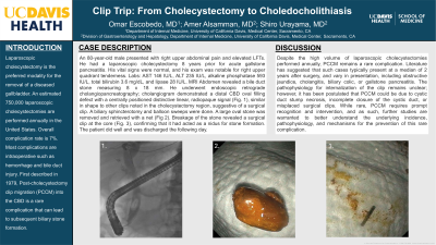
P3520 - Clip Trip: From Cholecystectomy to Choledocholithiasis
Tuesday, October 29, 2024
10:30 AM - 4:00 PM ET
Location: Exhibit Hall E

Has Audio
- OE
Omar Escobedo, MD
University of California Davis Health
Sacramento, CA
Presenting Author(s)
Omar Escobedo, MD, Amer Alsamman, MD, Shiro Urayama, MD
University of California Davis Health, Sacramento, CA
Introduction: Laparoscopic cholecystectomy is the preferred modality for the removal of a diseased gallbladder. An estimated 750,000 laparoscopic cholecystectomies are performed annually in the United States. Overall complication rate is 7%. Most complications are intraoperative such as hemorrhage and bile duct injury. First described in 1979, Post-cholecystectomy clip migration (PCCM) into the CBD is a rare complication that can lead to subsequent biliary stone formation
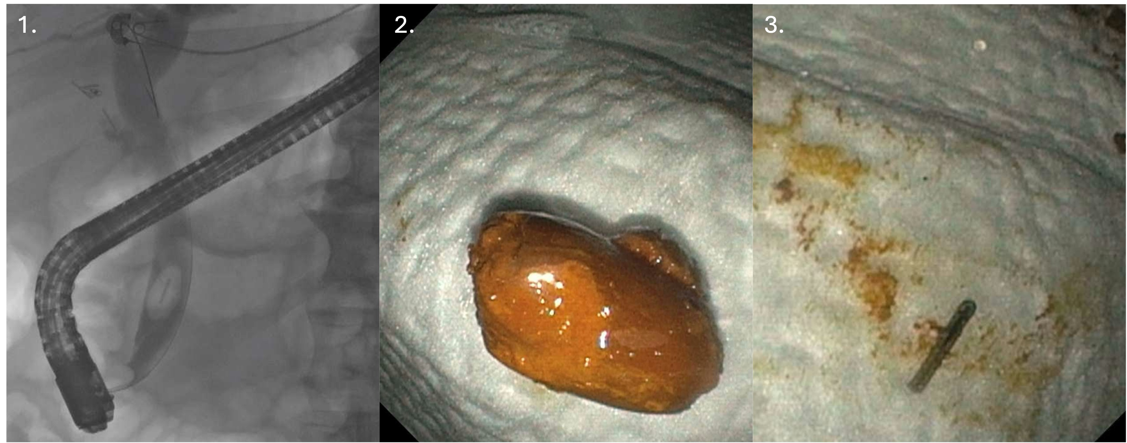
Case Description/Methods: An 80-year-old male presented with right upper abdominal pain and elevated LFTs. He had a laparoscopic cholecystectomy 8 years prior for acute gallstone pancreatitis. His vital signs were normal, and his exam was notable for right upper quadrant tenderness. Labs: AST 146 IU/L, ALT 235 IU/L, alkaline phosphatase 593 IU/L, total bilirubin 3.6 mg/dL, and lipase 28 IU/L. MRI Abdomen revealed a bile duct stone measuring 8 x 18 mm. He underwent endoscopic retrograde cholangiopancreatography; cholangiogram demonstrated a distal CBD oval filling defect with a centrally positioned distinctive linear, radiopaque signal (Fig. 1), similar in shape to other clips noted in the cholecystectomy region, suggestive of a surgical clip. A biliary sphincterotomy and balloon sweeps were done. A large oval stone was removed and retrieved with a net (Fig 2). Breakage of the stone revealed a surgical clip at the core (Fig. 3), confirming that it had acted as a nidus for stone formation. The patient did well and was discharged the following day.
Discussion: Despite the high volume of laparoscopic cholecystectomies performed annually, PCCM remains a rare complication. Literature has suggested that such cases typically present at a median of 2 years after surgery, and vary in presentation, including obstructive jaundice, cholangitis, biliary colic, or gallstone pancreatitis. The pathophysiology for internalization of the clip remains unclear; however, it has been postulated that PCCM could be due to cystic duct stump necrosis, incomplete closure of the cystic duct, or misplaced surgical clips. While rare, PCCM requires prompt recognition and intervention, and as such, further studies are warranted to better understand the underlying incidence, pathophysiology, and mechanisms for prevention of this rare complication.

Disclosures:
Omar Escobedo, MD, Amer Alsamman, MD, Shiro Urayama, MD. P3520 - Clip Trip: From Cholecystectomy to Choledocholithiasis, ACG 2024 Annual Scientific Meeting Abstracts. Philadelphia, PA: American College of Gastroenterology.
University of California Davis Health, Sacramento, CA
Introduction: Laparoscopic cholecystectomy is the preferred modality for the removal of a diseased gallbladder. An estimated 750,000 laparoscopic cholecystectomies are performed annually in the United States. Overall complication rate is 7%. Most complications are intraoperative such as hemorrhage and bile duct injury. First described in 1979, Post-cholecystectomy clip migration (PCCM) into the CBD is a rare complication that can lead to subsequent biliary stone formation
Case Description/Methods: An 80-year-old male presented with right upper abdominal pain and elevated LFTs. He had a laparoscopic cholecystectomy 8 years prior for acute gallstone pancreatitis. His vital signs were normal, and his exam was notable for right upper quadrant tenderness. Labs: AST 146 IU/L, ALT 235 IU/L, alkaline phosphatase 593 IU/L, total bilirubin 3.6 mg/dL, and lipase 28 IU/L. MRI Abdomen revealed a bile duct stone measuring 8 x 18 mm. He underwent endoscopic retrograde cholangiopancreatography; cholangiogram demonstrated a distal CBD oval filling defect with a centrally positioned distinctive linear, radiopaque signal (Fig. 1), similar in shape to other clips noted in the cholecystectomy region, suggestive of a surgical clip. A biliary sphincterotomy and balloon sweeps were done. A large oval stone was removed and retrieved with a net (Fig 2). Breakage of the stone revealed a surgical clip at the core (Fig. 3), confirming that it had acted as a nidus for stone formation. The patient did well and was discharged the following day.
Discussion: Despite the high volume of laparoscopic cholecystectomies performed annually, PCCM remains a rare complication. Literature has suggested that such cases typically present at a median of 2 years after surgery, and vary in presentation, including obstructive jaundice, cholangitis, biliary colic, or gallstone pancreatitis. The pathophysiology for internalization of the clip remains unclear; however, it has been postulated that PCCM could be due to cystic duct stump necrosis, incomplete closure of the cystic duct, or misplaced surgical clips. While rare, PCCM requires prompt recognition and intervention, and as such, further studies are warranted to better understand the underlying incidence, pathophysiology, and mechanisms for prevention of this rare complication.

Figure: Figure 1: A distal common bile duct filling defect with a central linear radiopaque signal.
Figure 2: Retrieved common bile duct stone.
Figure 3: A surgical clip retrieved from within the stone after crushing it.
Figure 2: Retrieved common bile duct stone.
Figure 3: A surgical clip retrieved from within the stone after crushing it.
Disclosures:
Omar Escobedo indicated no relevant financial relationships.
Amer Alsamman indicated no relevant financial relationships.
Shiro Urayama: Noah Medical – Consultant. Olympus America – Consultant.
Omar Escobedo, MD, Amer Alsamman, MD, Shiro Urayama, MD. P3520 - Clip Trip: From Cholecystectomy to Choledocholithiasis, ACG 2024 Annual Scientific Meeting Abstracts. Philadelphia, PA: American College of Gastroenterology.

