Monday Poster Session
Category: Stomach
P3421 - Isolated Gastric Outlet Obstruction Secondary to Metastatic Invasive Lobular Breast Cancer: A Case Report
Monday, October 28, 2024
10:30 AM - 4:00 PM ET
Location: Exhibit Hall E

Has Audio
- AJ
Amey Joshi, MD
Sparrow Hospital, Michigan State University
Lansing, MI
Presenting Author(s)
Amey Joshi, MD1, Hemangi Kale, MD2, Vishal Kaila, MD2
1Sparrow Hospital, Michigan State University, Lansing, MI; 2Baylor Scott & White Medical Center, Dallas, TX
Introduction: Gastric outlet obstruction (GOO) typically arises from mechanical causes impeding gastric emptying, including peptic ulcer disease and malignancy. While distal gastric and pancreatic cancers are well-documented to cause malignancy-related GOO, isolated GOO secondary to breast carcinoma is a rare occurrence. We report an unusual case of isolated GOO secondary to invasive lobular carcinoma (ILC) of the breast, initially suspected to be in remission, that was diagnosed with endoscopic ultrasound-guided fine needle aspiration (FNA) and treated with appropriate chemotherapy.
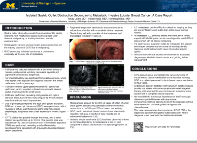
Case Description/Methods: A 69-year-old lady presented with reports of a two-week history of nausea and post-prandial vomiting. Her history was significant for ILC of the breast treated with adjuvant loco-regional radiation and lymph node resection four years prior. A double-contrast upper gastrointestinal series revealed a dilated stomach with slowed, partial emptying into the small bowel. EGD showed mild gastritis and pyloric stenosis without an intrinsic mass, and pyloric dilation was done. Due to persisting symptoms, EGD with endoscopic ultrasound was performed, which revealed diffuse wall thickening at the prepyloric region extending to the pylorus with a wall thickness of 14mm. FNA revealed poorly differentiated adenocarcinoma consistent with previously diagnosed ILC of the breast. The patient was started on targeted chemotherapy, and EUS repeated at one year of follow-up showed significant improvement in the appearance of antral wall thickness to 5.6mm with near resolution of patient symptoms.
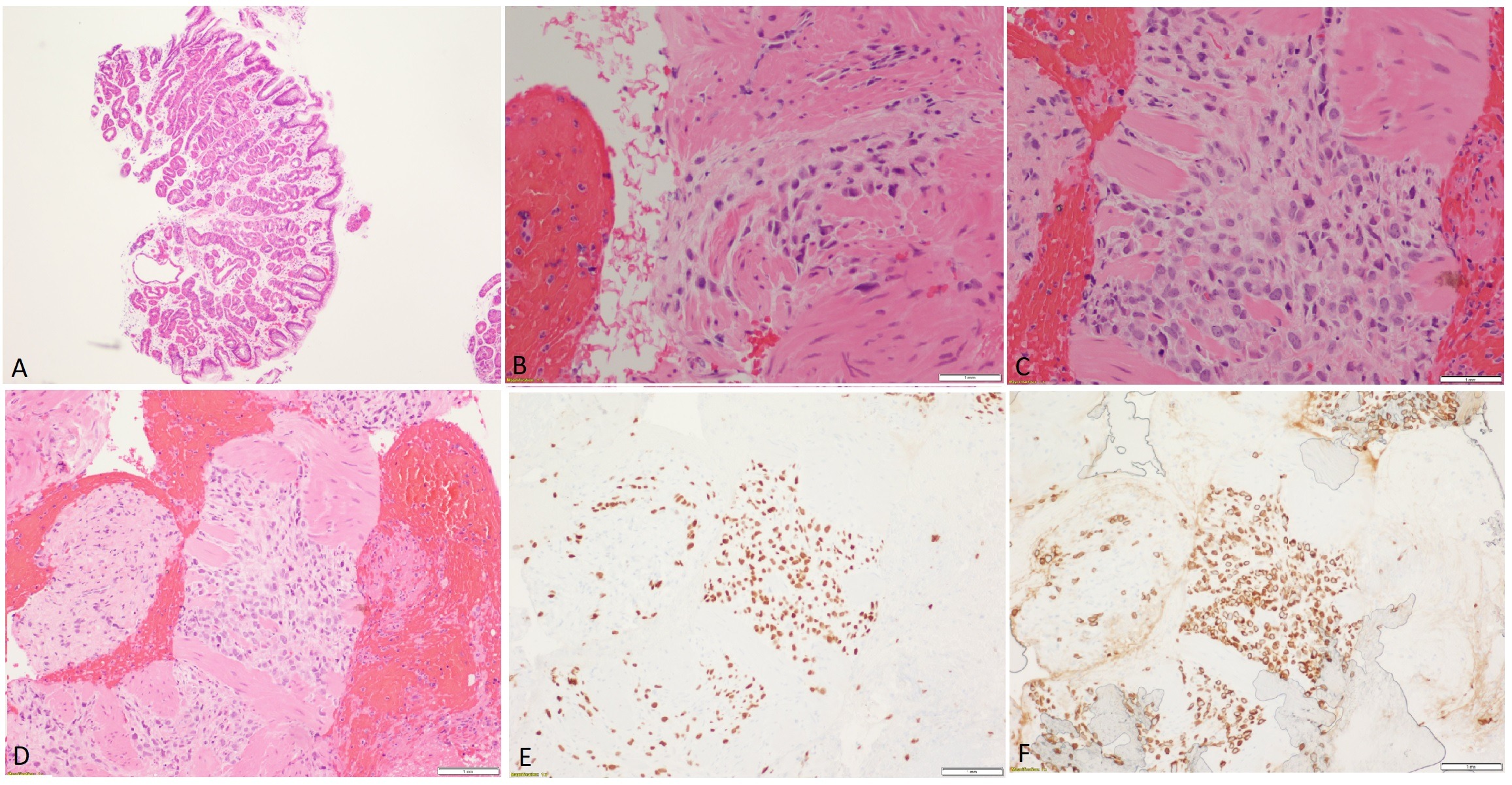
Discussion: Malignancies account for up to 80% of cases of GOO, of which obstruction due to metastatic breast cancer has been rarely reported, with only a handful of case reports. ILC has been observed to have an increased tendency to metastasize to the GI tract compared to other breast cancer subtypes. As metastatic ILC primarily affects the submucosal layers, superficial/initial biopsies can be non-diagnostic in 46-50% of cases, which can be falsely reassuring. Multiple biopsies may be crucial to making a timely diagnosis and initiating treatment of metastatic ILC with newer chemotherapeutic agents. This case demonstrates the importance of EUS FNA and appropriate immunohistochemical staining in the diagnostic algorithm for gastric outlet obstruction where the diagnosis is unclear with the traditional methods.

Disclosures:
Amey Joshi, MD1, Hemangi Kale, MD2, Vishal Kaila, MD2. P3421 - Isolated Gastric Outlet Obstruction Secondary to Metastatic Invasive Lobular Breast Cancer: A Case Report, ACG 2024 Annual Scientific Meeting Abstracts. Philadelphia, PA: American College of Gastroenterology.
1Sparrow Hospital, Michigan State University, Lansing, MI; 2Baylor Scott & White Medical Center, Dallas, TX
Introduction: Gastric outlet obstruction (GOO) typically arises from mechanical causes impeding gastric emptying, including peptic ulcer disease and malignancy. While distal gastric and pancreatic cancers are well-documented to cause malignancy-related GOO, isolated GOO secondary to breast carcinoma is a rare occurrence. We report an unusual case of isolated GOO secondary to invasive lobular carcinoma (ILC) of the breast, initially suspected to be in remission, that was diagnosed with endoscopic ultrasound-guided fine needle aspiration (FNA) and treated with appropriate chemotherapy.
Case Description/Methods: A 69-year-old lady presented with reports of a two-week history of nausea and post-prandial vomiting. Her history was significant for ILC of the breast treated with adjuvant loco-regional radiation and lymph node resection four years prior. A double-contrast upper gastrointestinal series revealed a dilated stomach with slowed, partial emptying into the small bowel. EGD showed mild gastritis and pyloric stenosis without an intrinsic mass, and pyloric dilation was done. Due to persisting symptoms, EGD with endoscopic ultrasound was performed, which revealed diffuse wall thickening at the prepyloric region extending to the pylorus with a wall thickness of 14mm. FNA revealed poorly differentiated adenocarcinoma consistent with previously diagnosed ILC of the breast. The patient was started on targeted chemotherapy, and EUS repeated at one year of follow-up showed significant improvement in the appearance of antral wall thickness to 5.6mm with near resolution of patient symptoms.
Discussion: Malignancies account for up to 80% of cases of GOO, of which obstruction due to metastatic breast cancer has been rarely reported, with only a handful of case reports. ILC has been observed to have an increased tendency to metastasize to the GI tract compared to other breast cancer subtypes. As metastatic ILC primarily affects the submucosal layers, superficial/initial biopsies can be non-diagnostic in 46-50% of cases, which can be falsely reassuring. Multiple biopsies may be crucial to making a timely diagnosis and initiating treatment of metastatic ILC with newer chemotherapeutic agents. This case demonstrates the importance of EUS FNA and appropriate immunohistochemical staining in the diagnostic algorithm for gastric outlet obstruction where the diagnosis is unclear with the traditional methods.

Figure: Histopathology of fine needle aspirated tissue in gastric tissue at the pylorus and gastric body A) Gastric body, B-D) Cell block, E) GATA 3 stain, and F) CK7 stain
Disclosures:
Amey Joshi indicated no relevant financial relationships.
Hemangi Kale indicated no relevant financial relationships.
Vishal Kaila indicated no relevant financial relationships.
Amey Joshi, MD1, Hemangi Kale, MD2, Vishal Kaila, MD2. P3421 - Isolated Gastric Outlet Obstruction Secondary to Metastatic Invasive Lobular Breast Cancer: A Case Report, ACG 2024 Annual Scientific Meeting Abstracts. Philadelphia, PA: American College of Gastroenterology.
