Monday Poster Session
Category: Stomach
P3422 - A Rare Case of Gastric Lymphangioma Masquerading as a Malignancy
Monday, October 28, 2024
10:30 AM - 4:00 PM ET
Location: Exhibit Hall E

Has Audio
- RP
Rahul Patel, DO
Jefferson Health
Washington Township, NJ
Presenting Author(s)
Award: Presidential Poster Award
Rahul Patel, DO1, Matthew Everwine, DO1, Brian Blair, DO2, C. Jonathan Foster, DO2
1Jefferson Health, Washington Township, NJ; 2Jefferson Health, Cherry Hill, NJ
Introduction: Cystic lymphangiomas are rare benign tumors. Typically, they occur in children in the neck or axilla. We present a gastric lymphangioma in which the patient presented with red flag symptoms.
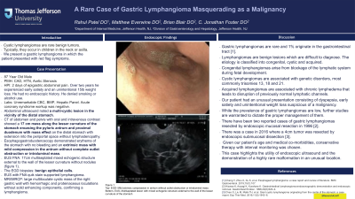
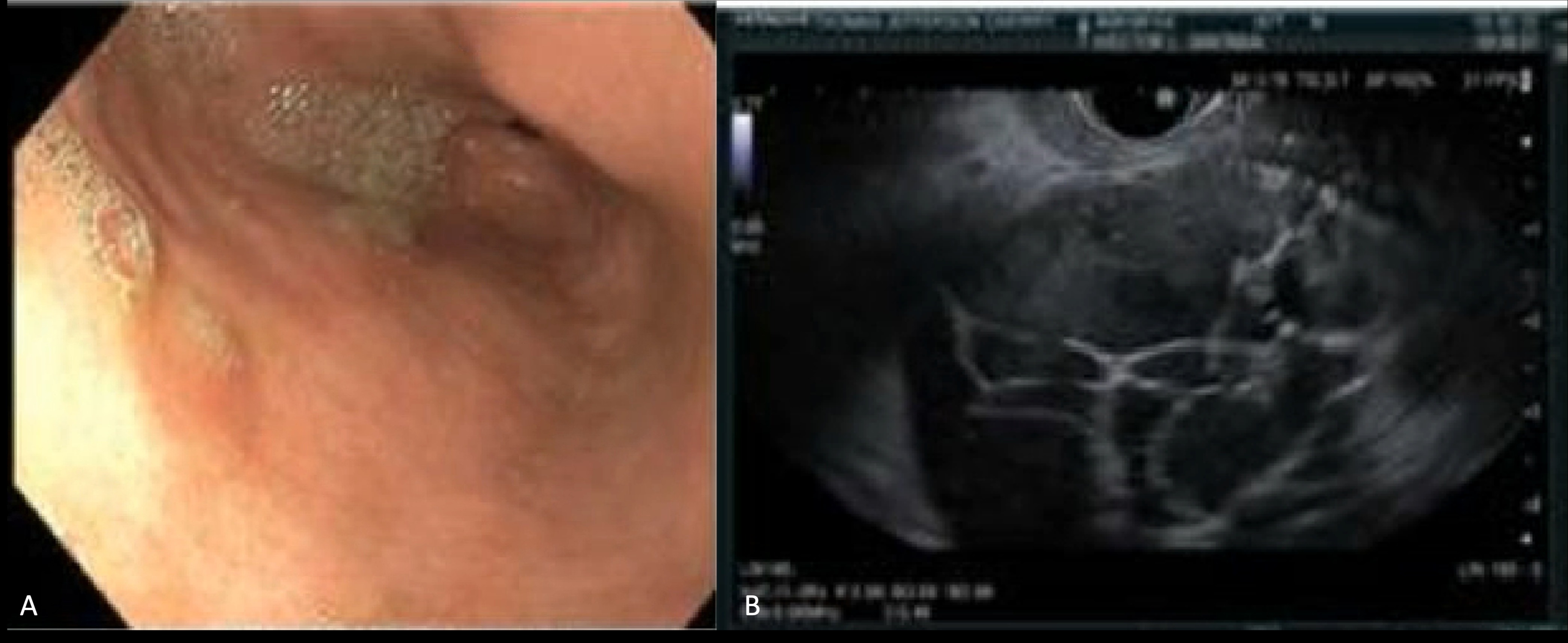
Case Description/Methods: An 87-year-old male with a history of coronary artery disease, hypertension and aortic stenosis presented with epigastric abdominal pain for two days. Over two years he experienced early satiety and an unintentional 15lb weight loss. He had no endoscopic history. He denied smoking or alcohol use. On arrival, he was hemodynamically stable and complete blood count, basic metabolic panel, hepatic panel, acute coronary syndrome workup was unremarkable. An abdominal ultrasound noted a multi-cystic lesion in the vicinity of the distal stomach. A computerized tomography (CT) of abdomen and pelvis with oral and intravenous contrast showed a 17 cm mass along the lesser curvature of the stomach encasing the pyloric antrum and proximal duodenum with mass effect on the distal stomach with extension into the periportal space without lymphadenopathy. He underwent an esophagogastroduodenoscopy (EGD) demonstrating erythema of the stomach with no bleeding and an extrinsic mass with mild compression in the antrum without complete outlet obstruction or intraluminal mass. 3 days later, he underwent an endoscopic ultrasound with fine needle aspiration (EUS FNA) showing a 17cm multiseptated mixed echogenic structure external to the wall of the lesser curvature without nodules (figure 1). The EGD biopsies demonstrated benign epithelial cells. EUS with FNA quik stain supported lymphangioma. Prior to discharge, a magnetic resonance cholangiopancreatography (MRCP) was performed, demonstrating a large multiloculate cystic mass of the right gastric wall with hemorrhagic and proteinaceous loculations without solid enhancing components, confirming a lymphangioma.
Discussion: Gastric lymphangiomas are rare and 1% originate in the gastrointestinal tract. Our patient had an unusual presentation consisting of dyspepsia, early satiety and unintentional weight loss suspicious of a malignancy. While there are no firm guidelines on gastric lymphangioma management due to its rarity, there have been cases of resections of them. Given our patient’s age and medical co-morbidities, conservative therapy with interval monitoring was chosen. This case highlights the utility of endoscopic ultrasound and the demonstration of a highly rare malformation in an unusual location.

Disclosures:
Rahul Patel, DO1, Matthew Everwine, DO1, Brian Blair, DO2, C. Jonathan Foster, DO2. P3422 - A Rare Case of Gastric Lymphangioma Masquerading as a Malignancy, ACG 2024 Annual Scientific Meeting Abstracts. Philadelphia, PA: American College of Gastroenterology.
Rahul Patel, DO1, Matthew Everwine, DO1, Brian Blair, DO2, C. Jonathan Foster, DO2
1Jefferson Health, Washington Township, NJ; 2Jefferson Health, Cherry Hill, NJ
Introduction: Cystic lymphangiomas are rare benign tumors. Typically, they occur in children in the neck or axilla. We present a gastric lymphangioma in which the patient presented with red flag symptoms.
Case Description/Methods: An 87-year-old male with a history of coronary artery disease, hypertension and aortic stenosis presented with epigastric abdominal pain for two days. Over two years he experienced early satiety and an unintentional 15lb weight loss. He had no endoscopic history. He denied smoking or alcohol use. On arrival, he was hemodynamically stable and complete blood count, basic metabolic panel, hepatic panel, acute coronary syndrome workup was unremarkable. An abdominal ultrasound noted a multi-cystic lesion in the vicinity of the distal stomach. A computerized tomography (CT) of abdomen and pelvis with oral and intravenous contrast showed a 17 cm mass along the lesser curvature of the stomach encasing the pyloric antrum and proximal duodenum with mass effect on the distal stomach with extension into the periportal space without lymphadenopathy. He underwent an esophagogastroduodenoscopy (EGD) demonstrating erythema of the stomach with no bleeding and an extrinsic mass with mild compression in the antrum without complete outlet obstruction or intraluminal mass. 3 days later, he underwent an endoscopic ultrasound with fine needle aspiration (EUS FNA) showing a 17cm multiseptated mixed echogenic structure external to the wall of the lesser curvature without nodules (figure 1). The EGD biopsies demonstrated benign epithelial cells. EUS with FNA quik stain supported lymphangioma. Prior to discharge, a magnetic resonance cholangiopancreatography (MRCP) was performed, demonstrating a large multiloculate cystic mass of the right gastric wall with hemorrhagic and proteinaceous loculations without solid enhancing components, confirming a lymphangioma.
Discussion: Gastric lymphangiomas are rare and 1% originate in the gastrointestinal tract. Our patient had an unusual presentation consisting of dyspepsia, early satiety and unintentional weight loss suspicious of a malignancy. While there are no firm guidelines on gastric lymphangioma management due to its rarity, there have been cases of resections of them. Given our patient’s age and medical co-morbidities, conservative therapy with interval monitoring was chosen. This case highlights the utility of endoscopic ultrasound and the demonstration of a highly rare malformation in an unusual location.

Figure: Figure 1
(A) EGD- Mild extrinsic compression in antrum without outlet obstruction or intraluminal mass
(B) EUS- Multiseptated lesion with mixed echogenic structure external to the wall of the lesser curvature of the stomach.
(A) EGD- Mild extrinsic compression in antrum without outlet obstruction or intraluminal mass
(B) EUS- Multiseptated lesion with mixed echogenic structure external to the wall of the lesser curvature of the stomach.
Disclosures:
Rahul Patel indicated no relevant financial relationships.
Matthew Everwine indicated no relevant financial relationships.
Brian Blair indicated no relevant financial relationships.
C. Jonathan Foster: Conmed – Consultant. Steris – Consultant.
Rahul Patel, DO1, Matthew Everwine, DO1, Brian Blair, DO2, C. Jonathan Foster, DO2. P3422 - A Rare Case of Gastric Lymphangioma Masquerading as a Malignancy, ACG 2024 Annual Scientific Meeting Abstracts. Philadelphia, PA: American College of Gastroenterology.

