Monday Poster Session
Category: Stomach
P3353 - A Rare Case of Gastric Leiomyoma Presenting With Acute Abdominal Pain
Monday, October 28, 2024
10:30 AM - 4:00 PM ET
Location: Exhibit Hall E

Has Audio
- TL
Tooba Laeeq, MD
University of Nevada
Las Vegas, NV
Presenting Author(s)
Tooba Laeeq, MD1, Amith Subhash, MD2, Shahid Wahid, MD2
1University of Nevada, Las Vegas, NV; 2Kirk Kerkorian School of Medicine at the University of Nevada, Las Vegas, NV
Introduction: Leiomyomas are rare tumors originating from smooth muscle cells. Gastric leiomyomas (GL) account for only 2.5% of gastric tumors, are usually asymptomatic, and are slow-growing. We present a case of symptomatic gastric leiomyoma.
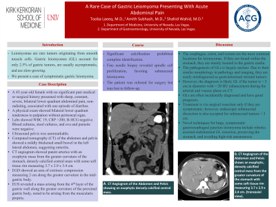
Case Description/Methods: A 43-year-old female with no significant past medical or surgical history presented with sharp, constant, severe, bilateral lower quadrant abdominal pain, non-radiating, associated with one episode of diarrhea. A physical exam showed bilateral lower quadrant tenderness to palpation without peritoneal signs. Labs showed WBC 19, CRP >200, B-HCG negative. Blood cultures, stool cultures, and ova and parasite were negative. Ultrasound pelvis was unremarkable. Computed tomography (CT) of the abdomen and pelvis showed a mildly thickened small bowel in the left lateral abdomen, suggesting enteritis. CT angiogram showed patent arteries with an exophytic mass from the greater curvature of the stomach, densely calcified central mass with some soft tissue rim measuring 3.7 x 2.9 x 3.4 cm. EGD showed an area of extrinsic compression measuring 2 cm along the greater curvature in the mid-gastric body. EUS revealed a mass arising from the 4th layer of the gastric wall along the greater curvature of the proximal gastric body, noted to be arising from the muscularis propria. Significant calcification prohibited complete identification. Fine needle biopsy revealed spindle cell proliferation, favoring submucosal leiomyoma. The patient was referred for surgery but was lost to follow-up.
Discussion: The esophagus, colon, and rectum are the most common locations for leiomyomas. If they are found within the stomach, they are mainly located in the gastric cardia. The pathogenesis of GLs is largely unclear. Due to the similarity in morphology in pathology and imaging, they are easily misdiagnosed as gastrointestinal stromal tumors. However, the diagnosis is likely GL if the tumor is < 5 cm in diameter with < 20 HU enhancement during the arterial and venous phase on CT. GLs are often incidentally diagnosed and have good prognosis. Treatment is via surgical resection only if they are symptomatic; however, endoscopic submucosal dissection is also accepted for submucosal tumors < 3 cm. Novel techniques for large, symptomatic gastroesophageal junction leiomyoma include robotic-assisted endoluminal GL resection, preserving the stomach, and avoiding high-risk anastomosis.

Disclosures:
Tooba Laeeq, MD1, Amith Subhash, MD2, Shahid Wahid, MD2. P3353 - A Rare Case of Gastric Leiomyoma Presenting With Acute Abdominal Pain, ACG 2024 Annual Scientific Meeting Abstracts. Philadelphia, PA: American College of Gastroenterology.
1University of Nevada, Las Vegas, NV; 2Kirk Kerkorian School of Medicine at the University of Nevada, Las Vegas, NV
Introduction: Leiomyomas are rare tumors originating from smooth muscle cells. Gastric leiomyomas (GL) account for only 2.5% of gastric tumors, are usually asymptomatic, and are slow-growing. We present a case of symptomatic gastric leiomyoma.
Case Description/Methods: A 43-year-old female with no significant past medical or surgical history presented with sharp, constant, severe, bilateral lower quadrant abdominal pain, non-radiating, associated with one episode of diarrhea. A physical exam showed bilateral lower quadrant tenderness to palpation without peritoneal signs. Labs showed WBC 19, CRP >200, B-HCG negative. Blood cultures, stool cultures, and ova and parasite were negative. Ultrasound pelvis was unremarkable. Computed tomography (CT) of the abdomen and pelvis showed a mildly thickened small bowel in the left lateral abdomen, suggesting enteritis. CT angiogram showed patent arteries with an exophytic mass from the greater curvature of the stomach, densely calcified central mass with some soft tissue rim measuring 3.7 x 2.9 x 3.4 cm. EGD showed an area of extrinsic compression measuring 2 cm along the greater curvature in the mid-gastric body. EUS revealed a mass arising from the 4th layer of the gastric wall along the greater curvature of the proximal gastric body, noted to be arising from the muscularis propria. Significant calcification prohibited complete identification. Fine needle biopsy revealed spindle cell proliferation, favoring submucosal leiomyoma. The patient was referred for surgery but was lost to follow-up.
Discussion: The esophagus, colon, and rectum are the most common locations for leiomyomas. If they are found within the stomach, they are mainly located in the gastric cardia. The pathogenesis of GLs is largely unclear. Due to the similarity in morphology in pathology and imaging, they are easily misdiagnosed as gastrointestinal stromal tumors. However, the diagnosis is likely GL if the tumor is < 5 cm in diameter with < 20 HU enhancement during the arterial and venous phase on CT. GLs are often incidentally diagnosed and have good prognosis. Treatment is via surgical resection only if they are symptomatic; however, endoscopic submucosal dissection is also accepted for submucosal tumors < 3 cm. Novel techniques for large, symptomatic gastroesophageal junction leiomyoma include robotic-assisted endoluminal GL resection, preserving the stomach, and avoiding high-risk anastomosis.

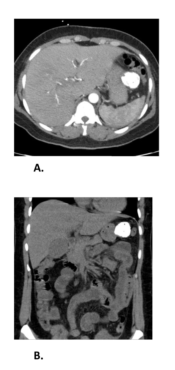
Figure: A. CT Angiogram of the Abdomen and Pelvis shows an exophytic, densely calcified, central mass from the greater curvature of the stomach with some soft tissue rim measuring 3.7 x 2.9 x 3.4 cm. (transaxial view).
B. CT Angiogram of the Abdomen and Pelvis (coronal view).
B. CT Angiogram of the Abdomen and Pelvis (coronal view).
Disclosures:
Tooba Laeeq indicated no relevant financial relationships.
Amith Subhash indicated no relevant financial relationships.
Shahid Wahid indicated no relevant financial relationships.
Tooba Laeeq, MD1, Amith Subhash, MD2, Shahid Wahid, MD2. P3353 - A Rare Case of Gastric Leiomyoma Presenting With Acute Abdominal Pain, ACG 2024 Annual Scientific Meeting Abstracts. Philadelphia, PA: American College of Gastroenterology.
