Monday Poster Session
Category: Stomach
P3365 - Recurrent Urothelial Cancer Presenting as Gastric Outlet Obstruction From Linitis Plastica, a Rare Manifestation
Monday, October 28, 2024
10:30 AM - 4:00 PM ET
Location: Exhibit Hall E

Has Audio

Raza Muhammad, DO
University of Chicago Northshore
Chicago, IL
Presenting Author(s)
Raza Muhammad, DO1, Edward Villa, MD2, Omar Abdelsadek, MD3
1University of Chicago Northshore, Evanston, IL; 2NorthShore University HealthSystem, Evanston, IL; 3NorthShore University HealthSystem, Chicago, IL
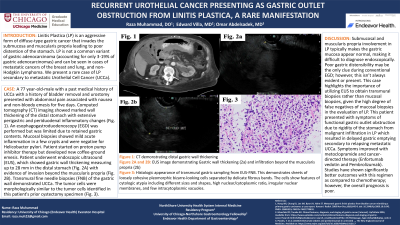
Introduction: Linitis Plastica (LP) is an aggressive form of diffuse-type gastric cancer that invades the submucosa and muscularis propria leading to poor distention of the stomach. LP is not a common variant of gastric adenocarcinoma (accounting for only 3-19% of gastric adenocarcinomas) and can be seen in cases of metastatic cancers of the breast and lung, and non-Hodgkin Lymphoma. We present a rare case of LP secondary to metastatic Urothelial Cell Cancer (UCCa).
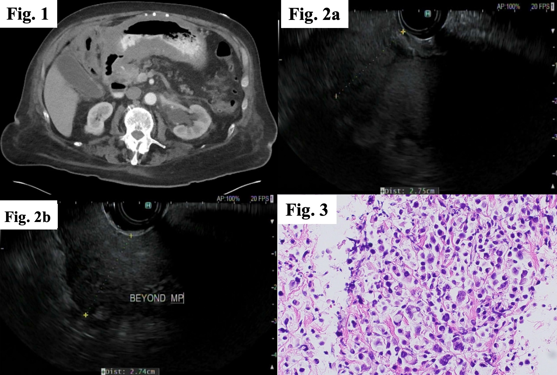
Case Description/Methods: A 77 year-old-male with a past medical history of UCCa with history of bladder removal and urostomy presented with abdominal pain associated with nausea and non-bloody emesis for five days. Computed tomography (CT) imaging showed marked wall thickening of the distal stomach with extensive perigastric and periduodenal inflammatory changes (Fig. 1). An esophagogastroduodenoscopy (EGD) was performed but was limited due to retained gastric contents. Mucosal biopsies showed mild acute inflammation in a few crypts and were negative for Helicobacter pylori. Patient started on proton pump inhibitor therapy but developed new coffee-ground emesis. Patient underwent endoscopic ultrasound (EUS), which showed gastric wall thickening measuring up to 28 mm in the distal stomach (Fig. 2A) with evidence of invasion beyond the muscularis propria (Fig. 2B). Transmural fine needle biopsies (FNB) of the gastric wall demonstrated UCCa. The tumor cells were morphologically similar to the tumor cells identified in this patient's prior cystectomy specimen (Fig. 3).
Discussion: Submucosal and muscularis propria involvement in LP typically makes the gastric mucosa appear normal, making it difficult to diagnose endoscopically. Poor gastric distensibility may be the only clue during conventional EGD; however, this isn’t always evident or present. This case highlights the importance of utilizing EUS to obtain transmural biopsies rather than mucosal biopsies, given the high degree of false negatives of mucosal biopsies in the evaluation of LP. This patient presented with symptoms of functional gastric outlet obstruction due to rigidity of the stomach from malignant infiltration in LP which resulted in delayed gastric emptying secondary to relapsing metastatic UCCa. Symptoms improved with metoclopramide and cancer-directed therapy (Enfortumab vedotin and Pembrolizumab). Studies have shown significantly better outcomes with this regimen as compared to chemotherapy; however, the overall prognosis is poor.

Disclosures:
Raza Muhammad, DO1, Edward Villa, MD2, Omar Abdelsadek, MD3. P3365 - Recurrent Urothelial Cancer Presenting as Gastric Outlet Obstruction From Linitis Plastica, a Rare Manifestation, ACG 2024 Annual Scientific Meeting Abstracts. Philadelphia, PA: American College of Gastroenterology.
1University of Chicago Northshore, Evanston, IL; 2NorthShore University HealthSystem, Evanston, IL; 3NorthShore University HealthSystem, Chicago, IL
Introduction: Linitis Plastica (LP) is an aggressive form of diffuse-type gastric cancer that invades the submucosa and muscularis propria leading to poor distention of the stomach. LP is not a common variant of gastric adenocarcinoma (accounting for only 3-19% of gastric adenocarcinomas) and can be seen in cases of metastatic cancers of the breast and lung, and non-Hodgkin Lymphoma. We present a rare case of LP secondary to metastatic Urothelial Cell Cancer (UCCa).
Case Description/Methods: A 77 year-old-male with a past medical history of UCCa with history of bladder removal and urostomy presented with abdominal pain associated with nausea and non-bloody emesis for five days. Computed tomography (CT) imaging showed marked wall thickening of the distal stomach with extensive perigastric and periduodenal inflammatory changes (Fig. 1). An esophagogastroduodenoscopy (EGD) was performed but was limited due to retained gastric contents. Mucosal biopsies showed mild acute inflammation in a few crypts and were negative for Helicobacter pylori. Patient started on proton pump inhibitor therapy but developed new coffee-ground emesis. Patient underwent endoscopic ultrasound (EUS), which showed gastric wall thickening measuring up to 28 mm in the distal stomach (Fig. 2A) with evidence of invasion beyond the muscularis propria (Fig. 2B). Transmural fine needle biopsies (FNB) of the gastric wall demonstrated UCCa. The tumor cells were morphologically similar to the tumor cells identified in this patient's prior cystectomy specimen (Fig. 3).
Discussion: Submucosal and muscularis propria involvement in LP typically makes the gastric mucosa appear normal, making it difficult to diagnose endoscopically. Poor gastric distensibility may be the only clue during conventional EGD; however, this isn’t always evident or present. This case highlights the importance of utilizing EUS to obtain transmural biopsies rather than mucosal biopsies, given the high degree of false negatives of mucosal biopsies in the evaluation of LP. This patient presented with symptoms of functional gastric outlet obstruction due to rigidity of the stomach from malignant infiltration in LP which resulted in delayed gastric emptying secondary to relapsing metastatic UCCa. Symptoms improved with metoclopramide and cancer-directed therapy (Enfortumab vedotin and Pembrolizumab). Studies have shown significantly better outcomes with this regimen as compared to chemotherapy; however, the overall prognosis is poor.

Figure: Figure 1: CT demonstrating distal gastric wall thickening
Figure 2A and 2B: EUS images demonstrating Gastric wall thickening (2A) and infiltration beyond the muscularis propria (2B)
Figure 3: Histologic appearance of transmural gastric sampling from EUS-FNB. This demonstrates sheets of loosely cohesive pleomorphic bizarre-looking cells separated by delicate fibrous bands. The cells show features of cytologic atypia including different sizes and shapes, high nuclear/cytoplasmic ratio, irregular nuclear membranes, and few intracytoplasmic vacuoles.
Disclosures:
Raza Muhammad indicated no relevant financial relationships.
Edward Villa: Interscope – Consultant. Olympus Corp – Consultant.
Omar Abdelsadek indicated no relevant financial relationships.
Raza Muhammad, DO1, Edward Villa, MD2, Omar Abdelsadek, MD3. P3365 - Recurrent Urothelial Cancer Presenting as Gastric Outlet Obstruction From Linitis Plastica, a Rare Manifestation, ACG 2024 Annual Scientific Meeting Abstracts. Philadelphia, PA: American College of Gastroenterology.
