Monday Poster Session
Category: Interventional Endoscopy
P2802 - Non-Invasive Approaches to Diagnosing Splenosis: A Case Report of Splenosis Mimicking a Pancreatic Mass
Monday, October 28, 2024
10:30 AM - 4:00 PM ET
Location: Exhibit Hall E

Has Audio

Mohamed Karam Douedari, MD
Mount Sinai Medical Center
Miami Beach, FL
Presenting Author(s)
Mohamed Karam Douedari, MD1, Rama Tarakji, MD1, Mahmoud Mahfouz, MD2
1Mount Sinai Medical Center, Miami Beach, FL; 2University of Miami Miller School of Medicine, Miami, FL
Introduction: Splenosis is a benign condition characterized by the heterotopic auto-transplantation of splenic tissue following splenic trauma or splenectomy, leading to the implantation of splenic tissue in various parts of the body. It is often asymptomatic and discovered incidentally. Recognizing splenosis is crucial as it can mimic malignant processes, thereby preventing unnecessary interventions, which can sometimes carry potential complications.
We present a case of a 59-year-old female with a history of splenectomy who was found to have a mass in the left upper quadrant initially suspected to be a pancreatic neoplasm. Further investigation with endoscopic ultrasound (EUS) and scintigraphy confirmed the diagnosis of splenosis.
Case Description/Methods: A 59-year-old female with a history of splenectomy performed 20 years ago following a motor vehicle accident who presented with back pain and chills.
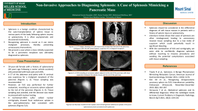
A CT of the abdomen and pelvis with IV contrast showed a 4.5 cm left upper quadrant soft tissue mass likely arising from the tail of the pancreas, suspicious for a malignant neoplasm. Tissue sampling was recommended. However, an EUS was performed for further evaluation, revealing an accessory spleen adjacent to the tail of the pancreas with a distinct separation between the pancreatic plane and the splenic tissue. Tissue sampling was avoided, and the decision was made to proceed with nuclear testing to confirm the diagnosis. A technetium-99m sulfur colloid nuclear scintigraphy showed focal radiotracer uptake in the post-splenectomy bed consistent with splenosis, correlating with the soft tissue density seen on CT.
Discussion: Splenosis should be considered in the differential diagnosis of soft tissue masses in patients with a history of splenic injury or splenectomy. Literature review shows that cases of splenosis are often misdiagnosed, leading to unnecessary invasive tissue sampling due to a low index of suspicion which could potentially result in significant bleeding. However, with the combination of EUS and scintigraphy, we were able to confidently diagnose splenosis without resorting to invasive procedures and avoiding the potential complications associated with tissue sampling.
References:
1. Tirado R et al., Splenosis: A Benign Phenomenon Mimicking Metastatic Cancer, American Journal of Gastroenterology, October 2015, 110(S): S176
2. Kim GE et al., Recognizing intrapancreatic accessory spleen via EUS: Interobserver variability, Endoscopic Ultrasound, November-December 2019, 8(6): 392-397

Disclosures:
Mohamed Karam Douedari, MD1, Rama Tarakji, MD1, Mahmoud Mahfouz, MD2. P2802 - Non-Invasive Approaches to Diagnosing Splenosis: A Case Report of Splenosis Mimicking a Pancreatic Mass, ACG 2024 Annual Scientific Meeting Abstracts. Philadelphia, PA: American College of Gastroenterology.
1Mount Sinai Medical Center, Miami Beach, FL; 2University of Miami Miller School of Medicine, Miami, FL
Introduction: Splenosis is a benign condition characterized by the heterotopic auto-transplantation of splenic tissue following splenic trauma or splenectomy, leading to the implantation of splenic tissue in various parts of the body. It is often asymptomatic and discovered incidentally. Recognizing splenosis is crucial as it can mimic malignant processes, thereby preventing unnecessary interventions, which can sometimes carry potential complications.
We present a case of a 59-year-old female with a history of splenectomy who was found to have a mass in the left upper quadrant initially suspected to be a pancreatic neoplasm. Further investigation with endoscopic ultrasound (EUS) and scintigraphy confirmed the diagnosis of splenosis.
Case Description/Methods: A 59-year-old female with a history of splenectomy performed 20 years ago following a motor vehicle accident who presented with back pain and chills.
A CT of the abdomen and pelvis with IV contrast showed a 4.5 cm left upper quadrant soft tissue mass likely arising from the tail of the pancreas, suspicious for a malignant neoplasm. Tissue sampling was recommended. However, an EUS was performed for further evaluation, revealing an accessory spleen adjacent to the tail of the pancreas with a distinct separation between the pancreatic plane and the splenic tissue. Tissue sampling was avoided, and the decision was made to proceed with nuclear testing to confirm the diagnosis. A technetium-99m sulfur colloid nuclear scintigraphy showed focal radiotracer uptake in the post-splenectomy bed consistent with splenosis, correlating with the soft tissue density seen on CT.
Discussion: Splenosis should be considered in the differential diagnosis of soft tissue masses in patients with a history of splenic injury or splenectomy. Literature review shows that cases of splenosis are often misdiagnosed, leading to unnecessary invasive tissue sampling due to a low index of suspicion which could potentially result in significant bleeding. However, with the combination of EUS and scintigraphy, we were able to confidently diagnose splenosis without resorting to invasive procedures and avoiding the potential complications associated with tissue sampling.
References:
1. Tirado R et al., Splenosis: A Benign Phenomenon Mimicking Metastatic Cancer, American Journal of Gastroenterology, October 2015, 110(S): S176
2. Kim GE et al., Recognizing intrapancreatic accessory spleen via EUS: Interobserver variability, Endoscopic Ultrasound, November-December 2019, 8(6): 392-397

Figure: Figures 1-2: CT images showing a 4.5 cm left upper quadrant soft tissue mass arising from the tail of the pancreas, contiguous with the left adrenal gland, suspicious for a malignant neoplasm.
Figures 3-4: EUS images revealing an accessory spleen adjacent to the tail of the pancreas.
Figures 5-6: technetium-99m sulfur colloid nuclear scintigraphy showing focal radiotracer uptake in the post-splenectomy bed consistent with splenosis
Figures 3-4: EUS images revealing an accessory spleen adjacent to the tail of the pancreas.
Figures 5-6: technetium-99m sulfur colloid nuclear scintigraphy showing focal radiotracer uptake in the post-splenectomy bed consistent with splenosis
Disclosures:
Mohamed Karam Douedari indicated no relevant financial relationships.
Rama Tarakji indicated no relevant financial relationships.
Mahmoud Mahfouz indicated no relevant financial relationships.
Mohamed Karam Douedari, MD1, Rama Tarakji, MD1, Mahmoud Mahfouz, MD2. P2802 - Non-Invasive Approaches to Diagnosing Splenosis: A Case Report of Splenosis Mimicking a Pancreatic Mass, ACG 2024 Annual Scientific Meeting Abstracts. Philadelphia, PA: American College of Gastroenterology.
