Monday Poster Session
Category: Interventional Endoscopy
P2808 - Endoscopic Ultrasound-Guided Fine-Needle Biopsy Unveils a Rare Mediastinal Ectopic Thyroid in an Elderly Male
Monday, October 28, 2024
10:30 AM - 4:00 PM ET
Location: Exhibit Hall E

Has Audio

Dimo Dimitrov, MD
St. Elizabeth's Medical Center, Boston University School of Medicine
Boston, MA
Presenting Author(s)
Dimo Dimitrov, MD1, Jad Mitri, MD1, Arpit Jain, MD1, Virginia Litle, MD1, Sandeep Krishnan, MD, PhD2
1St. Elizabeth's Medical Center, Boston University School of Medicine, Boston, MA; 2St. Elizabeth's Medical Center, Boston University School of Medicine, Brighton, MA
Introduction: Mediastinal masses pose significant diagnostic challenges due to multiple possible etiologies, ectopic thyroid being one of them. Very rarely, it can appear in the mediastinum and comprises < 1% of all mediastinal masses. This report highlights the rare case of ectopic thyroid diagnosed by endoscopic ultrasound (EUS). We discuss the diagnostic approach and examine the role of EUS-guided biopsy in evaluating mediastinal masses, providing insights for managing similar cases.
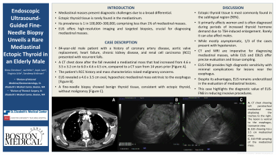
Case Description/Methods: An 84-year-old male with a complex medical history, including coronary artery disease, aortic valve replacement, heart failure, a previously known mediastinal mass, chronic kidney disease, renal cell carcinoma (RCC), presented with recurrent falls. CT chest with contrast revealed a mediastinal mass, which had increased in size from 4.6 x 3.5 x 3.2 cm to 6.0 x 4.6 x 4.5 cm over a period of 14 years. The mass displaced the trachea without obstructing it and extended between the trachea and esophagus. The patient's history of RCC and the characteristics of the enlarging mediastinal mass raised concerns for malignancy. He was referred to thoracic surgery for evaluation, and to a gastroenterologist for EUS for tissue diagnosis. EUS revealed an oval, hypoechoic mass in the mediastinum, extrinsic to the esophagus, measuring 4.6 x 3.5 cm. A fine-needle biopsy was performed without complications. Pathology results showed benign-appearing thyroid tissue, with no malignancy, consistent with ectopic thyroid tissue.
Discussion: This case highlights the importance of considering a broad differential when evaluating mediastinal solid masses. Ectopic thyroid tissue is more common in females and is usually diagnosed during periods of increased thyroid hormone demand such as menopause. However, it can have an atypical presentation, such as in our elderly male patient in his 80s. CT and MRI are imperative for diagnosing mediastinal masses, while EUS and endobronchial ultrasound (EBUS) offer precise evaluation and tissue sampling opportunities. EUS-guided fine-needle biopsy (EUS-FNB) is particularly effective for detailed imaging and biopsies of lesions adjacent to the esophagus, providing high diagnostic sensitivity with minimal complications. Despite its advantages, EUS remains underutilized in the evaluation of thoracic pathology. Our case demonstrates the significant diagnostic value and potential of EUS-FNB in reducing the need for more invasive procedures.

Disclosures:
Dimo Dimitrov, MD1, Jad Mitri, MD1, Arpit Jain, MD1, Virginia Litle, MD1, Sandeep Krishnan, MD, PhD2. P2808 - Endoscopic Ultrasound-Guided Fine-Needle Biopsy Unveils a Rare Mediastinal Ectopic Thyroid in an Elderly Male, ACG 2024 Annual Scientific Meeting Abstracts. Philadelphia, PA: American College of Gastroenterology.
1St. Elizabeth's Medical Center, Boston University School of Medicine, Boston, MA; 2St. Elizabeth's Medical Center, Boston University School of Medicine, Brighton, MA
Introduction: Mediastinal masses pose significant diagnostic challenges due to multiple possible etiologies, ectopic thyroid being one of them. Very rarely, it can appear in the mediastinum and comprises < 1% of all mediastinal masses. This report highlights the rare case of ectopic thyroid diagnosed by endoscopic ultrasound (EUS). We discuss the diagnostic approach and examine the role of EUS-guided biopsy in evaluating mediastinal masses, providing insights for managing similar cases.
Case Description/Methods: An 84-year-old male with a complex medical history, including coronary artery disease, aortic valve replacement, heart failure, a previously known mediastinal mass, chronic kidney disease, renal cell carcinoma (RCC), presented with recurrent falls. CT chest with contrast revealed a mediastinal mass, which had increased in size from 4.6 x 3.5 x 3.2 cm to 6.0 x 4.6 x 4.5 cm over a period of 14 years. The mass displaced the trachea without obstructing it and extended between the trachea and esophagus. The patient's history of RCC and the characteristics of the enlarging mediastinal mass raised concerns for malignancy. He was referred to thoracic surgery for evaluation, and to a gastroenterologist for EUS for tissue diagnosis. EUS revealed an oval, hypoechoic mass in the mediastinum, extrinsic to the esophagus, measuring 4.6 x 3.5 cm. A fine-needle biopsy was performed without complications. Pathology results showed benign-appearing thyroid tissue, with no malignancy, consistent with ectopic thyroid tissue.
Discussion: This case highlights the importance of considering a broad differential when evaluating mediastinal solid masses. Ectopic thyroid tissue is more common in females and is usually diagnosed during periods of increased thyroid hormone demand such as menopause. However, it can have an atypical presentation, such as in our elderly male patient in his 80s. CT and MRI are imperative for diagnosing mediastinal masses, while EUS and endobronchial ultrasound (EBUS) offer precise evaluation and tissue sampling opportunities. EUS-guided fine-needle biopsy (EUS-FNB) is particularly effective for detailed imaging and biopsies of lesions adjacent to the esophagus, providing high diagnostic sensitivity with minimal complications. Despite its advantages, EUS remains underutilized in the evaluation of thoracic pathology. Our case demonstrates the significant diagnostic value and potential of EUS-FNB in reducing the need for more invasive procedures.

Figure: A. CT chest demonstrating left paratracheal mediastinal mass displacing the trachea to the right. The lesion is ventral to the esophagus, transverse view. | B. EUS demonstrating 4.6 x 3.5 cm mediastinal mass. | C. EUS-FNB sampling of the mediastinal mass.
Disclosures:
Dimo Dimitrov indicated no relevant financial relationships.
Jad Mitri indicated no relevant financial relationships.
Arpit Jain indicated no relevant financial relationships.
Virginia Litle indicated no relevant financial relationships.
Sandeep Krishnan indicated no relevant financial relationships.
Dimo Dimitrov, MD1, Jad Mitri, MD1, Arpit Jain, MD1, Virginia Litle, MD1, Sandeep Krishnan, MD, PhD2. P2808 - Endoscopic Ultrasound-Guided Fine-Needle Biopsy Unveils a Rare Mediastinal Ectopic Thyroid in an Elderly Male, ACG 2024 Annual Scientific Meeting Abstracts. Philadelphia, PA: American College of Gastroenterology.
