Monday Poster Session
Category: IBD
P2716 - Inflammatory Pseudotumor: A Rare Presentation of Crohn's Disease
Monday, October 28, 2024
10:30 AM - 4:00 PM ET
Location: Exhibit Hall E

Has Audio

Carla Barberan Parraga, MD
Maimonides Medical Center
New York, NY
Presenting Author(s)
Carla Barberan Parraga, MD1, Samantha S. Ehrlich, MD2, Steve Obanor, MD2, Meredith E. Pittman, MD2, Manol Jovani, MD2
1Maimonides Medical Center, New York, NY; 2Maimonides Medical Center, Brooklyn, NY
Introduction: Crohn's disease (CD) is an inflammatory bowel disease that affects the entire gastrointestinal tract and often is located in the terminal ileum. It presents with vague symptoms like diarrhea, abdominal pain, and weight loss or complications due to transmural inflammation, such as fistulas, strictures, abscesses, and perianal involvement. Inflammatory pseudotumors (IPT) are well-defined, non-encapsulated, rare, quasi-neoplastic lesions originating from the unregulated growth of inflammatory cells. They can originate in any organ, but gastrointestinal tract involvement is rare.
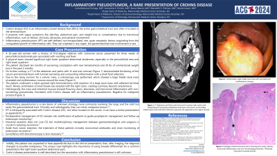
Case Description/Methods: We present a case of a 62-year-old woman who presented to the emergency department because of three weeks of periumbilical abdominal pain associated with vomiting and fevers. On examination, the pain was significant in the right lower abdominal quadrant. The patient endorsed a history of worsening constipation with hematochezia for the preceding 10 months which was associated with an unintentional weight loss of 30 lbs in the last 3 months. The patient has a significant family history of colon cancer her daughter and father. A computed tomography (CT) of the abdomen with IV and oral contrast demonstrated thickening of the cecum and terminal ileum with luminal narrowing and surrounding inflammation and a small pericecal collection. There was no evidence of obstruction. At colonoscopy a large friable cecal mass with ulcerated and erythematous mucosa was seen. At robot-assisted right hemicolectomy a large cecal mass with adherent terminal ileum was seen. Ninety centimeters of small bowel was resected along with the right colon with primary ileocolic anastomosis. Histologically, the mass and surrounding intestinal mucosa showed fissuring ulcers, abscesses, transmural inflammation, and non-necrotizing granulomata. The pathologic features were consistent with Crohn’s disease with an inflammatory pseudotumor, negative for malignancy.
Discussion: Inflammatory pseudotumors are rare and have infrequently been associated with Crohn’s disease. This case highlights a unique initial presentation of Crohn’s disease. Postoperative management of CD includes assessment of risk factors in order to guide management with anti-inflammatory therapy. The treatment of these patients often includes monoclonal antibodies and close monitoring of endoscopic recurrence. Surveillance with ileocolonoscopy is also necessary.

Disclosures:
Carla Barberan Parraga, MD1, Samantha S. Ehrlich, MD2, Steve Obanor, MD2, Meredith E. Pittman, MD2, Manol Jovani, MD2. P2716 - Inflammatory Pseudotumor: A Rare Presentation of Crohn's Disease, ACG 2024 Annual Scientific Meeting Abstracts. Philadelphia, PA: American College of Gastroenterology.
1Maimonides Medical Center, New York, NY; 2Maimonides Medical Center, Brooklyn, NY
Introduction: Crohn's disease (CD) is an inflammatory bowel disease that affects the entire gastrointestinal tract and often is located in the terminal ileum. It presents with vague symptoms like diarrhea, abdominal pain, and weight loss or complications due to transmural inflammation, such as fistulas, strictures, abscesses, and perianal involvement. Inflammatory pseudotumors (IPT) are well-defined, non-encapsulated, rare, quasi-neoplastic lesions originating from the unregulated growth of inflammatory cells. They can originate in any organ, but gastrointestinal tract involvement is rare.
Case Description/Methods: We present a case of a 62-year-old woman who presented to the emergency department because of three weeks of periumbilical abdominal pain associated with vomiting and fevers. On examination, the pain was significant in the right lower abdominal quadrant. The patient endorsed a history of worsening constipation with hematochezia for the preceding 10 months which was associated with an unintentional weight loss of 30 lbs in the last 3 months. The patient has a significant family history of colon cancer her daughter and father. A computed tomography (CT) of the abdomen with IV and oral contrast demonstrated thickening of the cecum and terminal ileum with luminal narrowing and surrounding inflammation and a small pericecal collection. There was no evidence of obstruction. At colonoscopy a large friable cecal mass with ulcerated and erythematous mucosa was seen. At robot-assisted right hemicolectomy a large cecal mass with adherent terminal ileum was seen. Ninety centimeters of small bowel was resected along with the right colon with primary ileocolic anastomosis. Histologically, the mass and surrounding intestinal mucosa showed fissuring ulcers, abscesses, transmural inflammation, and non-necrotizing granulomata. The pathologic features were consistent with Crohn’s disease with an inflammatory pseudotumor, negative for malignancy.
Discussion: Inflammatory pseudotumors are rare and have infrequently been associated with Crohn’s disease. This case highlights a unique initial presentation of Crohn’s disease. Postoperative management of CD includes assessment of risk factors in order to guide management with anti-inflammatory therapy. The treatment of these patients often includes monoclonal antibodies and close monitoring of endoscopic recurrence. Surveillance with ileocolonoscopy is also necessary.

Figure: A.Colonoscopy. Large friable cecal mass with ulcerated and erythematous mucosa.
B.Histology. Intestinal mucosa with a fissuring ulcer (blue star), abscess, transmural inflammation, and non-necrotizing granulomata. Thickened muscularis mucosae (green arrow). Most consistent with Crohns disease with inflammatory pseudotumor.
C.CT scan with PO and IV contrast. Severe wall thickening and narrowing of distal/terminal ileum and cecum. Surrounding inflammation and small fluid collection representing appendix matted to this region.
Disclosures:
Carla Barberan Parraga indicated no relevant financial relationships.
Samantha Ehrlich indicated no relevant financial relationships.
Steve Obanor indicated no relevant financial relationships.
Meredith Pittman indicated no relevant financial relationships.
Manol Jovani indicated no relevant financial relationships.
Carla Barberan Parraga, MD1, Samantha S. Ehrlich, MD2, Steve Obanor, MD2, Meredith E. Pittman, MD2, Manol Jovani, MD2. P2716 - Inflammatory Pseudotumor: A Rare Presentation of Crohn's Disease, ACG 2024 Annual Scientific Meeting Abstracts. Philadelphia, PA: American College of Gastroenterology.
