Monday Poster Session
Category: IBD
P2730 - Not Every Terminal Ileitis is Crohn's Disease: A Case of IGA Vasculitis-Associated Terminal Ileitis
Monday, October 28, 2024
10:30 AM - 4:00 PM ET
Location: Exhibit Hall E

Has Audio
- YR
Yasir Rajwana, MD
Marshall University Joan C. Edwards School of Medicine
Huntington, WV
Presenting Author(s)
Yasir Rajwana, MD1, Muhammad Rahoma saad, MD2, Ahmed Sherif, MD1
1Marshall University Joan C. Edwards School of Medicine, Huntington, WV; 2Marshall University, Huntington, WV
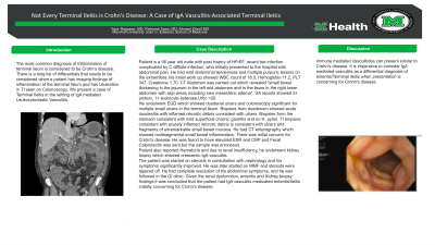
Introduction: The most common diagnosis of inflammation of terminal ileitis is considered to be Crohn's disease. There is a long list of differentials that needs to be considered when a patient has imaging findings of inflammation of the terminal Ileum and has Ulceration in TI seen on Colonoscopy. We present a case of Terminal Ileitis in the setting of IgA mediated Leukocytoclastic Vasculitis.
Case Description/Methods: Patient is a 56 year old male with past history of HFrEF, recent toe infection complicated by C difficile infection, who initially presented to the hospital with abdominal pain. He had mild abdominal tenderness and multiple purpuric lesions on the extremities.His initial work up showed WBC count of 18.3, Hemoglobin 11.2, PLT 347, Creatinine 1.70. CT Abdomen was carried out which revealed "small bowel thickening in the jejunum in the left mid abdomen and in the ileum in the right lower abdomen with skip areas including new mesenteric edema". UA reports showed 2+ protein, 1+ leukocyte esterase,Urbc >20.
He underwent EGD which showed duodenal ulcers and colonoscopy significant for multiple small ulcers in the terminal ileum. Biopsies from duodenum showed acute duodenitis with inflamed necrotic debris consistent with ulcers. Biopsies from the stomach consistent with mild superficial chronic gastritis and no H. pylori. TI biopsies consistent with acutely inflamed necrotic debris is consistent with ulcers and fragments of unremarkable small bowel mucosa. He had CT enterography which showed multisegmental small bowel inflammation. There was initial concern for Crohn's disease. He was found to have elevated ESR and CRP and Fecal Calprotectin was sent but the sample was erroneous.
Patient also reported Hematuria and due to renal insufficiency, he underwent kidney biopsy which showed crescentic IgA vasculitis.
The patient was started on steroids in consultation with nephrology and his symptoms significantly improved. He was later started on MMF and steroids were tapered off. He had complete resolution of his abdominal symptoms, and he was followed in the GI clinic. Given the renal dysfunction, enteritis and Kidney biopsy findings it was concluded that the patient had IgA vasculitis medicated enteritis/Ileitis initially concerning for Crohn's disease.
Discussion: Immune mediated Vasculitides can present similar to Crohn's disease. It is imperative to consider IgA mediated vasculitis as a differential diagnosis of enteritis/Terminal ileitis when presentation is concerning for Crohn's disease.
Disclosures:
Yasir Rajwana, MD1, Muhammad Rahoma saad, MD2, Ahmed Sherif, MD1. P2730 - Not Every Terminal Ileitis is Crohn's Disease: A Case of IGA Vasculitis-Associated Terminal Ileitis, ACG 2024 Annual Scientific Meeting Abstracts. Philadelphia, PA: American College of Gastroenterology.
1Marshall University Joan C. Edwards School of Medicine, Huntington, WV; 2Marshall University, Huntington, WV
Introduction: The most common diagnosis of inflammation of terminal ileitis is considered to be Crohn's disease. There is a long list of differentials that needs to be considered when a patient has imaging findings of inflammation of the terminal Ileum and has Ulceration in TI seen on Colonoscopy. We present a case of Terminal Ileitis in the setting of IgA mediated Leukocytoclastic Vasculitis.
Case Description/Methods: Patient is a 56 year old male with past history of HFrEF, recent toe infection complicated by C difficile infection, who initially presented to the hospital with abdominal pain. He had mild abdominal tenderness and multiple purpuric lesions on the extremities.His initial work up showed WBC count of 18.3, Hemoglobin 11.2, PLT 347, Creatinine 1.70. CT Abdomen was carried out which revealed "small bowel thickening in the jejunum in the left mid abdomen and in the ileum in the right lower abdomen with skip areas including new mesenteric edema". UA reports showed 2+ protein, 1+ leukocyte esterase,Urbc >20.
He underwent EGD which showed duodenal ulcers and colonoscopy significant for multiple small ulcers in the terminal ileum. Biopsies from duodenum showed acute duodenitis with inflamed necrotic debris consistent with ulcers. Biopsies from the stomach consistent with mild superficial chronic gastritis and no H. pylori. TI biopsies consistent with acutely inflamed necrotic debris is consistent with ulcers and fragments of unremarkable small bowel mucosa. He had CT enterography which showed multisegmental small bowel inflammation. There was initial concern for Crohn's disease. He was found to have elevated ESR and CRP and Fecal Calprotectin was sent but the sample was erroneous.
Patient also reported Hematuria and due to renal insufficiency, he underwent kidney biopsy which showed crescentic IgA vasculitis.
The patient was started on steroids in consultation with nephrology and his symptoms significantly improved. He was later started on MMF and steroids were tapered off. He had complete resolution of his abdominal symptoms, and he was followed in the GI clinic. Given the renal dysfunction, enteritis and Kidney biopsy findings it was concluded that the patient had IgA vasculitis medicated enteritis/Ileitis initially concerning for Crohn's disease.
Discussion: Immune mediated Vasculitides can present similar to Crohn's disease. It is imperative to consider IgA mediated vasculitis as a differential diagnosis of enteritis/Terminal ileitis when presentation is concerning for Crohn's disease.
Disclosures:
Yasir Rajwana indicated no relevant financial relationships.
Muhammad Rahoma saad indicated no relevant financial relationships.
Ahmed Sherif indicated no relevant financial relationships.
Yasir Rajwana, MD1, Muhammad Rahoma saad, MD2, Ahmed Sherif, MD1. P2730 - Not Every Terminal Ileitis is Crohn's Disease: A Case of IGA Vasculitis-Associated Terminal Ileitis, ACG 2024 Annual Scientific Meeting Abstracts. Philadelphia, PA: American College of Gastroenterology.
