Monday Poster Session
Category: GI Bleeding
P2518 - Massive Upper Gastrointestinal Bleeding Following Transesophageal Echocardiography
Monday, October 28, 2024
10:30 AM - 4:00 PM ET
Location: Exhibit Hall E

Has Audio
- EA
Elvis J. Arteaga, MD
University of Arizona College of Medicine
Phoenix, AZ
Presenting Author(s)
Elvis J. Arteaga, MD1, Brian M.. Fung, MD2, Joseph David, MD1
1University of Arizona College of Medicine, Phoenix, AZ; 2Arizona Digestive Health, Mesa, AZ
Introduction: Transesophageal echocardiography (TEE) is a frequently used technique to examine the heart and great vessels that involves the advancement of an ultrasound probe into the esophagus. The procedure is relatively safe with reported rates of gastrointestinal (GI) bleeding and GI tear/perforation of 0.17% and 0.05%, respectively. In the following case, we describe a case of massive bleeding from esophageal injury during TEE.
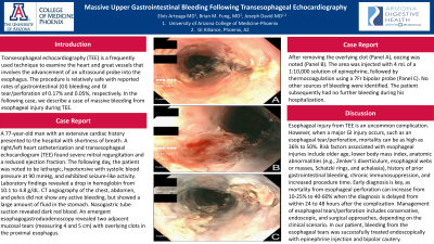
Case Description/Methods: A 77-year-old man with an extensive cardiac history presented to the hospital with shortness of breath. A right/left heart catheterization and transesophageal echocardiogram (TEE) found severe mitral regurgitation and a reduced ejection fraction. The following day, the patient was noted to be lethargic, hypotensive with systolic blood pressure at 90 mmHg, and exhibited seizure-like activity. Laboratory findings revealed a drop in hemoglobin from 10.1 to 4.8 g/dL. CT angiography of the chest, abdomen, and pelvis did not show any active bleeding, but showed a large amount of fluid in the stomach. Nasogastric tube suction revealed dark red blood. An emergent esophagogastroduodenoscopy revealed two adjacent mucosal tears (measuring 4 and 5 cm) with overlying clots in the proximal esophagus. After removing the overlying clot, oozing was noted. The area was injected with 4 mL of a 1:10,000 solution of epinephrine, followed by thermocoagulation using a 7Fr bipolar probe. No other sources of bleeding were identified. The patient subsequently had no further bleeding during his hospitalization.
Discussion: Esophageal injury from TEE is an uncommon complication. However, when a major GI injury occurs, such as an esophageal tear/perforation, mortality can be as high as 36% to 50%. Risk factors associated with esophageal injuries include older age, lower body mass index, anatomic abnormalities (e.g., Zenker’s diverticulum, esophageal webs or masses, Schatzki rings, and achalasia), history of prior gastrointestinal bleeding, chronic immunosuppression, and increased procedure time. Early diagnosis is key, as mortality from esophageal perforation can increase from 10-25% to 40-60% when the diagnosis is delayed from within 24 to 48 hours after the complication. Management of esophageal tears/perforation includes conservative, endoscopic, and surgical approaches, depending on the clinical scenario. In our patient, bleeding from the esophageal tears was successfully treated endoscopically with epinephrine injection and bipolar cautery.

Disclosures:
Elvis J. Arteaga, MD1, Brian M.. Fung, MD2, Joseph David, MD1. P2518 - Massive Upper Gastrointestinal Bleeding Following Transesophageal Echocardiography, ACG 2024 Annual Scientific Meeting Abstracts. Philadelphia, PA: American College of Gastroenterology.
1University of Arizona College of Medicine, Phoenix, AZ; 2Arizona Digestive Health, Mesa, AZ
Introduction: Transesophageal echocardiography (TEE) is a frequently used technique to examine the heart and great vessels that involves the advancement of an ultrasound probe into the esophagus. The procedure is relatively safe with reported rates of gastrointestinal (GI) bleeding and GI tear/perforation of 0.17% and 0.05%, respectively. In the following case, we describe a case of massive bleeding from esophageal injury during TEE.
Case Description/Methods: A 77-year-old man with an extensive cardiac history presented to the hospital with shortness of breath. A right/left heart catheterization and transesophageal echocardiogram (TEE) found severe mitral regurgitation and a reduced ejection fraction. The following day, the patient was noted to be lethargic, hypotensive with systolic blood pressure at 90 mmHg, and exhibited seizure-like activity. Laboratory findings revealed a drop in hemoglobin from 10.1 to 4.8 g/dL. CT angiography of the chest, abdomen, and pelvis did not show any active bleeding, but showed a large amount of fluid in the stomach. Nasogastric tube suction revealed dark red blood. An emergent esophagogastroduodenoscopy revealed two adjacent mucosal tears (measuring 4 and 5 cm) with overlying clots in the proximal esophagus. After removing the overlying clot, oozing was noted. The area was injected with 4 mL of a 1:10,000 solution of epinephrine, followed by thermocoagulation using a 7Fr bipolar probe. No other sources of bleeding were identified. The patient subsequently had no further bleeding during his hospitalization.
Discussion: Esophageal injury from TEE is an uncommon complication. However, when a major GI injury occurs, such as an esophageal tear/perforation, mortality can be as high as 36% to 50%. Risk factors associated with esophageal injuries include older age, lower body mass index, anatomic abnormalities (e.g., Zenker’s diverticulum, esophageal webs or masses, Schatzki rings, and achalasia), history of prior gastrointestinal bleeding, chronic immunosuppression, and increased procedure time. Early diagnosis is key, as mortality from esophageal perforation can increase from 10-25% to 40-60% when the diagnosis is delayed from within 24 to 48 hours after the complication. Management of esophageal tears/perforation includes conservative, endoscopic, and surgical approaches, depending on the clinical scenario. In our patient, bleeding from the esophageal tears was successfully treated endoscopically with epinephrine injection and bipolar cautery.

Figure: A. Esophageal tears with thrombus, B. Esophageal tear after clot removal. C. Esophageal tears after cauterization.
Disclosures:
Elvis Arteaga indicated no relevant financial relationships.
Brian Fung indicated no relevant financial relationships.
Joseph David indicated no relevant financial relationships.
Elvis J. Arteaga, MD1, Brian M.. Fung, MD2, Joseph David, MD1. P2518 - Massive Upper Gastrointestinal Bleeding Following Transesophageal Echocardiography, ACG 2024 Annual Scientific Meeting Abstracts. Philadelphia, PA: American College of Gastroenterology.
