Monday Poster Session
Category: Esophagus
P2311 - A Doomsday Bacteria or an Innocent Bystander: Sarcina ventriculi Associated Esophagitis - A Case Report
Monday, October 28, 2024
10:30 AM - 4:00 PM ET
Location: Exhibit Hall E

Has Audio
.jpg)
Irene J. Lee, MD
Baylor College of Medicine
Houston, TX
Presenting Author(s)
Irene J. Lee, MD, Stalin R. Subramanian, MD, Shipla Jain, MD
Baylor College of Medicine, Houston, TX
Introduction: While esophagitis is often secondary to chronic acid reflux disease and can be managed in the outpatient setting, we must always consider more treacherous pathologies requiring targeted treatment. We present a case of odynophagia secondary to Sarcina ventriculi infection of the esophagus. Although our patient recovered on an antibiotic regimen, treatment was delayed due to the rarity of Sarcina ventriculi esophagitis and the lack of diagnostic and management guidelines. The recognition of this infectious pathogen has been increasing in recent decades, yet its consequence to humans remains unknown due to its low prevalence.
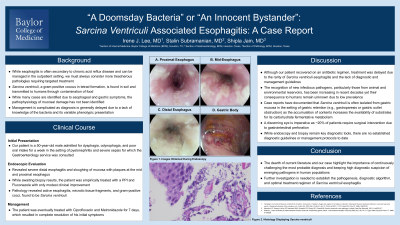
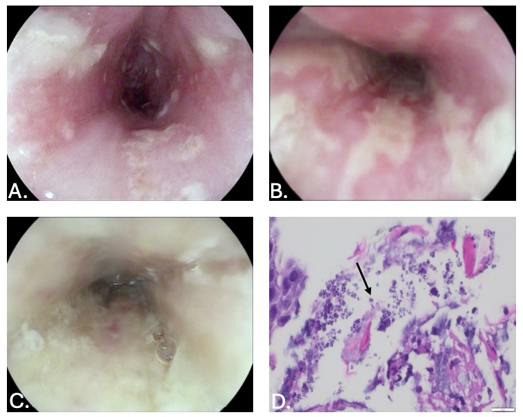
Case Description/Methods: Our patient is a 50-year-old male admitted for dysphagia, odynophagia, and poor oral intake for a week in the setting of pyelonephritis and severe sepsis for which the Gastroenterology service was consulted. Endoscopy revealed severe distal esophagitis and sloughing of mucosa with plaques at the mid and proximal esophagus. While awaiting biopsy results, the patient was empirically treated with a PPI and fluconazole with only modest clinical improvement. Pathology revealed active esophagitis, necrotic tissue fragments, and gram-positive cocci, found to be Sarcina ventriculi. The patient was eventually treated with Ciprofloxacin and Metronidazole for 7 days, which resulted in complete resolution of his initial symptoms.
Discussion: Sarcina ventriculi, a gram-positive coccus in tetrad formation, is found in soil and transmitted to humans through contamination of food. Case reports have documented that this pathogen is often isolated from gastric mucosa in the setting of gastric retention (e.g., gastroparesis or gastric outlet obstruction) as the accumulation of contents increases the availability of substrates for its carbohydrate fermentative metabolism; however, the pathophysiology of mucosal damage is unknown. Management is complicated as diagnosis is often delayed due to a lack of knowledge of the bacteria and its variable phenotypic presentation. However, a discerning eye is imperative since ~20% of patients require surgical intervention due to gastrointestinal perforation.
The dearth of current literature and our case highlight the importance of continuously challenging the most probable diagnosis and keeping high diagnostic suspicion of emerging pathogens in human populations. Further investigation is needed to establish the pathogenesis, diagnostic algorithm, and treatment of Sarcina ventriculi esophagitis.

Disclosures:
Irene J. Lee, MD, Stalin R. Subramanian, MD, Shipla Jain, MD. P2311 - A Doomsday Bacteria or an Innocent Bystander: <i>Sarcina ventriculi</i> Associated Esophagitis - A Case Report, ACG 2024 Annual Scientific Meeting Abstracts. Philadelphia, PA: American College of Gastroenterology.
Baylor College of Medicine, Houston, TX
Introduction: While esophagitis is often secondary to chronic acid reflux disease and can be managed in the outpatient setting, we must always consider more treacherous pathologies requiring targeted treatment. We present a case of odynophagia secondary to Sarcina ventriculi infection of the esophagus. Although our patient recovered on an antibiotic regimen, treatment was delayed due to the rarity of Sarcina ventriculi esophagitis and the lack of diagnostic and management guidelines. The recognition of this infectious pathogen has been increasing in recent decades, yet its consequence to humans remains unknown due to its low prevalence.
Case Description/Methods: Our patient is a 50-year-old male admitted for dysphagia, odynophagia, and poor oral intake for a week in the setting of pyelonephritis and severe sepsis for which the Gastroenterology service was consulted. Endoscopy revealed severe distal esophagitis and sloughing of mucosa with plaques at the mid and proximal esophagus. While awaiting biopsy results, the patient was empirically treated with a PPI and fluconazole with only modest clinical improvement. Pathology revealed active esophagitis, necrotic tissue fragments, and gram-positive cocci, found to be Sarcina ventriculi. The patient was eventually treated with Ciprofloxacin and Metronidazole for 7 days, which resulted in complete resolution of his initial symptoms.
Discussion: Sarcina ventriculi, a gram-positive coccus in tetrad formation, is found in soil and transmitted to humans through contamination of food. Case reports have documented that this pathogen is often isolated from gastric mucosa in the setting of gastric retention (e.g., gastroparesis or gastric outlet obstruction) as the accumulation of contents increases the availability of substrates for its carbohydrate fermentative metabolism; however, the pathophysiology of mucosal damage is unknown. Management is complicated as diagnosis is often delayed due to a lack of knowledge of the bacteria and its variable phenotypic presentation. However, a discerning eye is imperative since ~20% of patients require surgical intervention due to gastrointestinal perforation.
The dearth of current literature and our case highlight the importance of continuously challenging the most probable diagnosis and keeping high diagnostic suspicion of emerging pathogens in human populations. Further investigation is needed to establish the pathogenesis, diagnostic algorithm, and treatment of Sarcina ventriculi esophagitis.

Figure: Figure 1. Images Obtained During Endoscopy and Biopsy. A - proximal esophagus, B - mid-esophagus, C - distal esophagus, D - histology displaying Sarcina ventriculi
Disclosures:
Irene Lee indicated no relevant financial relationships.
Stalin Subramanian indicated no relevant financial relationships.
Shipla Jain indicated no relevant financial relationships.
Irene J. Lee, MD, Stalin R. Subramanian, MD, Shipla Jain, MD. P2311 - A Doomsday Bacteria or an Innocent Bystander: <i>Sarcina ventriculi</i> Associated Esophagitis - A Case Report, ACG 2024 Annual Scientific Meeting Abstracts. Philadelphia, PA: American College of Gastroenterology.
