Monday Poster Session
Category: Esophagus
P2320 - Concurrent Rare Parahiatal Hernia With a Sliding Hiatal Hernia Seen on Esophagogastroduodenoscopy: A Case Report
Monday, October 28, 2024
10:30 AM - 4:00 PM ET
Location: Exhibit Hall E

Has Audio
.jpg)
Aarohi Parikh, MD
HCA Healthcare
Kingwood, TX
Presenting Author(s)
Aarohi Parikh, MD, Anam Zara, MD, Ismail Hader, MD
HCA Healthcare, Kingwood, TX
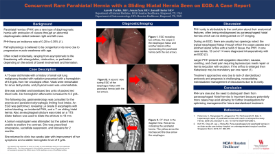
Introduction: Parahiatal hernias (PHH) are rare diaphragmatic hernias between the left and right crus with an incidence of 0.20 to 0.35%1. They are often asymptomatic and incidentally noted through radiology, endoscopy, or surgery. However, PHH can present with severe abdominal pain if complicated by incarceration or obstruction. We describe a rare case of PHH and a sliding hiatal hernia (SHH) in a female who underwent an esophagogastroduodenoscopy (EGD).
Case Description/Methods: A 73-year old female with a history of small cell lung cancer treated with radiation presented with a hemoglobin of 6.8 g/dL. Vitals were remarkable for sinus tachycardia, and physical exam was unremarkable. She was admitted and was transfused one unit of packed red blood cells. The following day, gastroenterology was consulted for anemia and persistent odynophagia limiting food intake. An EGD revealed LA Grade D esophagitis with active bleeding, an incidental PHH, and a 7 cm SHH. A barium esophagram was attempted but the patient was unable to swallow the contrast. She was prescribed omeprazole, sucralfate suspension, and lidocaine for two months. She returned to clinic two weeks later, with improvement of her symptoms and a stable hemoglobin level. The biopsy of the gastric tissue was negative for Helicobacter pylori.
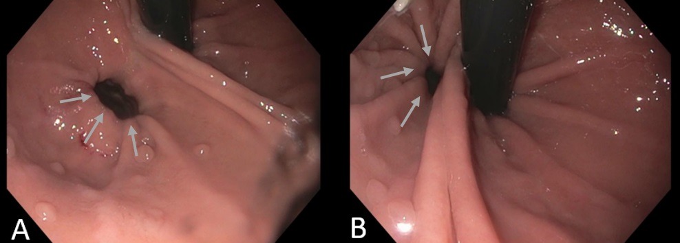
Discussion: PHH are uncommon due to their anatomical features, often being misdiagnosed as paraesophageal HH. The preferred diagnostic tool is a CT scan of the chest or abdomen to properly distinguish the anatomy. During an EGD, two openings are visible: the typical esophageal hiatus through which the scope passes and another later orifice with a band of tissue, the PHH (Figure 1). Larger PHH present with epigastric discomfort, nausea, vomiting, and chest pain requiring laparoscopic mesh repair or hernia reduction with excision. Treatment approaches vary due to lack of standardized protocols and prognosis is challenging, necessitating individualized management discussions due to its rarity.
Reference:
1. Palanivelu C, Rangarajan M, Jategaonkar PA, Parthasarathi R, Balu K. Laparoscopic repair of parahiatal hernias with mesh: a retrospective study. Hernia. 2008 Oct;12(5):521-5. doi: 10.1007/s10029-008-0380-2.

Disclosures:
Aarohi Parikh, MD, Anam Zara, MD, Ismail Hader, MD. P2320 - Concurrent Rare Parahiatal Hernia With a Sliding Hiatal Hernia Seen on Esophagogastroduodenoscopy: A Case Report, ACG 2024 Annual Scientific Meeting Abstracts. Philadelphia, PA: American College of Gastroenterology.
HCA Healthcare, Kingwood, TX
Introduction: Parahiatal hernias (PHH) are rare diaphragmatic hernias between the left and right crus with an incidence of 0.20 to 0.35%1. They are often asymptomatic and incidentally noted through radiology, endoscopy, or surgery. However, PHH can present with severe abdominal pain if complicated by incarceration or obstruction. We describe a rare case of PHH and a sliding hiatal hernia (SHH) in a female who underwent an esophagogastroduodenoscopy (EGD).
Case Description/Methods: A 73-year old female with a history of small cell lung cancer treated with radiation presented with a hemoglobin of 6.8 g/dL. Vitals were remarkable for sinus tachycardia, and physical exam was unremarkable. She was admitted and was transfused one unit of packed red blood cells. The following day, gastroenterology was consulted for anemia and persistent odynophagia limiting food intake. An EGD revealed LA Grade D esophagitis with active bleeding, an incidental PHH, and a 7 cm SHH. A barium esophagram was attempted but the patient was unable to swallow the contrast. She was prescribed omeprazole, sucralfate suspension, and lidocaine for two months. She returned to clinic two weeks later, with improvement of her symptoms and a stable hemoglobin level. The biopsy of the gastric tissue was negative for Helicobacter pylori.
Discussion: PHH are uncommon due to their anatomical features, often being misdiagnosed as paraesophageal HH. The preferred diagnostic tool is a CT scan of the chest or abdomen to properly distinguish the anatomy. During an EGD, two openings are visible: the typical esophageal hiatus through which the scope passes and another later orifice with a band of tissue, the PHH (Figure 1). Larger PHH present with epigastric discomfort, nausea, vomiting, and chest pain requiring laparoscopic mesh repair or hernia reduction with excision. Treatment approaches vary due to lack of standardized protocols and prognosis is challenging, necessitating individualized management discussions due to its rarity.
Reference:
1. Palanivelu C, Rangarajan M, Jategaonkar PA, Parthasarathi R, Balu K. Laparoscopic repair of parahiatal hernias with mesh: a retrospective study. Hernia. 2008 Oct;12(5):521-5. doi: 10.1007/s10029-008-0380-2.

Figure: Figure 1. A. EGD revealing two orifices, the scope in the esophagus hiatus and another lateral orifice representing the parahiatal hernia (grey arrows). B. A second view during EGD of the esophagus hiatus with parahiatal hernia (grey arrows).
Disclosures:
Aarohi Parikh indicated no relevant financial relationships.
Anam Zara indicated no relevant financial relationships.
Ismail Hader indicated no relevant financial relationships.
Aarohi Parikh, MD, Anam Zara, MD, Ismail Hader, MD. P2320 - Concurrent Rare Parahiatal Hernia With a Sliding Hiatal Hernia Seen on Esophagogastroduodenoscopy: A Case Report, ACG 2024 Annual Scientific Meeting Abstracts. Philadelphia, PA: American College of Gastroenterology.
