Monday Poster Session
Category: Colon
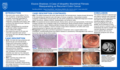
P2060 - Elusive Shadows: A Case of Idiopathic Myointimal Fibrosis Masquerading as Recurrent Colon Cancer
Monday, October 28, 2024
10:30 AM - 4:00 PM ET
Location: Exhibit Hall E

Has Audio

Parul Berry, MBBS
Mayo Clinic
Rochester, MN
Presenting Author(s)
Parul Berry, MBBS, Raseen Tariq, MBBS, Sahil Khanna, MBBS, MS, Conor Loftus, MD
Mayo Clinic, Rochester, MN
Introduction: The incidence of recurrent colon cancer varies from 14-21%, depending on the location of the primary tumor. We present a challenging case of a patient with a history of colon cancer, presenting with abdominal pain and hematochezia.
Case Description/Methods: A 50-year-old male with stage 2 colon carcinoma, treated with left hemicolectomy and primary anastomosis 6 years ago presented with lower abdominal pain, and watery diarrhea with occasional hematochezia for two weeks. On presentation, a computed tomography scan of the abdomen and pelvis revealed increased mural hyperenhancement in the sigmoid colon, thickening of the sigmoid and descending colon walls, and irregular wall thickening in the rectum, indeterminate for recurrent neoplasm.
The patient was admitted, and a colonoscopy was performed to rule out recurrent malignancy. The gastrointestinal pathogen panel was negative. Vitals remained stable during admission.
Colonoscopy showed congested, erythematous, and friable mucosa in the sigmoid colon. Biopsies revealed colonic mucosa with superficial erosions, lamina propria vascular congestion, and regenerative epithelial changes without dysplasia. Anticoagulation was deemed non-beneficial due to chronic congestion.
Post-discharge patient experienced recurrence of symptoms. Chronic inferior mesenteric vein occlusion secondary to malignancy and previous bowel resection was considered. Due to ongoing symptoms and lack of utility of medical therapy, a referral to colorectal surgery for resection of the congested segment was made.
The constellation of symptoms, imaging and endoscopic findings were suggestive of idiopathic myointimal hyperplasia of mesenteric veins. A curative extensive resection to allow a tension-free anastomosis was considered, given the patient’s previous surgery. Surgical options included a trial anastomosis versus proctectomy with end colostomy. After shared decision-making with the patient, it was decided to proceed with a laparoscopic, possibly open, low anterior resection with colorectal anastomosis. Three weeks post-surgery, the patient remained stable.
Discussion: This case highlights the complexity of idiopathic myointimal hyperplasia of mesenteric veins, a rare diagnosis presenting with progressive abdominal pain, and bloody diarrhea. Friability and congestion on endoscopy and intimal hyperproliferation on histopathology is seen.It often mimics inflammatory bowel disease and, in this case, colon cancer recurrence, posing significant diagnostic and therapeutic challenges.

Disclosures:
Parul Berry, MBBS, Raseen Tariq, MBBS, Sahil Khanna, MBBS, MS, Conor Loftus, MD. P2060 - Elusive Shadows: A Case of Idiopathic Myointimal Fibrosis Masquerading as Recurrent Colon Cancer, ACG 2024 Annual Scientific Meeting Abstracts. Philadelphia, PA: American College of Gastroenterology.
Mayo Clinic, Rochester, MN
Introduction: The incidence of recurrent colon cancer varies from 14-21%, depending on the location of the primary tumor. We present a challenging case of a patient with a history of colon cancer, presenting with abdominal pain and hematochezia.
Case Description/Methods: A 50-year-old male with stage 2 colon carcinoma, treated with left hemicolectomy and primary anastomosis 6 years ago presented with lower abdominal pain, and watery diarrhea with occasional hematochezia for two weeks. On presentation, a computed tomography scan of the abdomen and pelvis revealed increased mural hyperenhancement in the sigmoid colon, thickening of the sigmoid and descending colon walls, and irregular wall thickening in the rectum, indeterminate for recurrent neoplasm.
The patient was admitted, and a colonoscopy was performed to rule out recurrent malignancy. The gastrointestinal pathogen panel was negative. Vitals remained stable during admission.
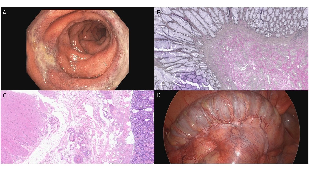
Colonoscopy showed congested, erythematous, and friable mucosa in the sigmoid colon. Biopsies revealed colonic mucosa with superficial erosions, lamina propria vascular congestion, and regenerative epithelial changes without dysplasia. Anticoagulation was deemed non-beneficial due to chronic congestion.
Post-discharge patient experienced recurrence of symptoms. Chronic inferior mesenteric vein occlusion secondary to malignancy and previous bowel resection was considered. Due to ongoing symptoms and lack of utility of medical therapy, a referral to colorectal surgery for resection of the congested segment was made.
The constellation of symptoms, imaging and endoscopic findings were suggestive of idiopathic myointimal hyperplasia of mesenteric veins. A curative extensive resection to allow a tension-free anastomosis was considered, given the patient’s previous surgery. Surgical options included a trial anastomosis versus proctectomy with end colostomy. After shared decision-making with the patient, it was decided to proceed with a laparoscopic, possibly open, low anterior resection with colorectal anastomosis. Three weeks post-surgery, the patient remained stable.
Discussion: This case highlights the complexity of idiopathic myointimal hyperplasia of mesenteric veins, a rare diagnosis presenting with progressive abdominal pain, and bloody diarrhea. Friability and congestion on endoscopy and intimal hyperproliferation on histopathology is seen.It often mimics inflammatory bowel disease and, in this case, colon cancer recurrence, posing significant diagnostic and therapeutic challenges.

Figure: Image A: Colonoscopy showing congested, erythematous and friable mucosa in sigmoid colon.
Image B: Verfoeff-van Gieson stained Histopathological slide showing intimal hyperplasia of transmural vessels and mucosal vessels thickening.
Image C: Histopathological slide showing intimal hyperplasia in transmural vessels.
Image D: Surgical Resection of 29 cm of Sigmoid colon
Image B: Verfoeff-van Gieson stained Histopathological slide showing intimal hyperplasia of transmural vessels and mucosal vessels thickening.
Image C: Histopathological slide showing intimal hyperplasia in transmural vessels.
Image D: Surgical Resection of 29 cm of Sigmoid colon
Disclosures:
Parul Berry indicated no relevant financial relationships.
Raseen Tariq indicated no relevant financial relationships.
Sahil Khanna: Ferring Pharmaceuticals, Inc. – Grant/Research Support. Finch – Grant/Research Support. Pfizer – Grant/Research Support. Probio Tech, LLC – Consultant. Rise – Consultant. Seres Therapeutics – Grant/Research Support. Takeda – Consultant. Vedanta – Grant/Research Support.
Conor Loftus indicated no relevant financial relationships.
Parul Berry, MBBS, Raseen Tariq, MBBS, Sahil Khanna, MBBS, MS, Conor Loftus, MD. P2060 - Elusive Shadows: A Case of Idiopathic Myointimal Fibrosis Masquerading as Recurrent Colon Cancer, ACG 2024 Annual Scientific Meeting Abstracts. Philadelphia, PA: American College of Gastroenterology.
