Monday Poster Session
Category: Colon
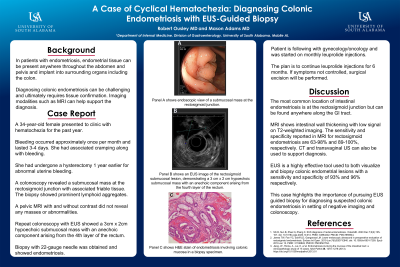
P2019 - A Case of Cyclical Hematochezia: Diagnosing Colonic Endometriosis With EUS-Guided Biopsy
Monday, October 28, 2024
10:30 AM - 4:00 PM ET
Location: Exhibit Hall E

Has Audio
- RO
Robert Ousley, MD
University of South Alabama
Mobile, AL
Presenting Author(s)
Robert Ousley, MD1, Mason G. Adams, MD2
1University of South Alabama, Mobile, AL; 2University of South Alabama College of Medicine, Mobile, AL
Introduction: Endometriosis is the presence of endometrial glands and stroma outside the uterus. Endometrial tissue can be present anywhere throughout the pelvis and abdomen and can implant into surrounding organs. Sub-peritoneal invasion by lesions exceeding 5 mm is considered deep endometriosis (DE). Endometriosis can be found in multiple locations, including the bowel. We present a case of colonic endometriosis diagnosed by EUS-guided fine needle biopsy.
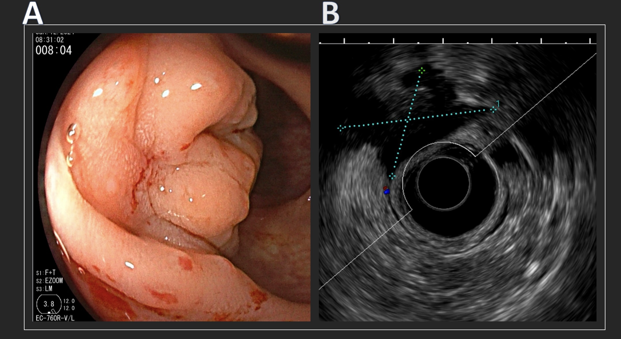
Case Description/Methods: A 34-year-old female presented to the gastroenterology clinic with hematochezia. Her bleeding was cyclical in nature and associated with abdominal cramps, lasting approximately 3-4 days and recurring monthly. The patient did not have menstrual cycles, having undergone a hysterectomy for abnormal uterine bleeding one year prior. Initial lab assessment in the clinic revealed mild anemia with a hemoglobin level of 11.2 g/dL. A colonoscopy revealed a submucosal mass at the rectosigmoid junction with associated friable tissue (Figure 1A). Biopsy showed colonic mucosa with prominent lymphoid aggregates. An MRI pelvis with and without contrast did not show any masses or abnormalities. The patient underwent a repeat colonoscopy with EUS, which showed a 3 cm x 2 cm hypoechoic submucosal mass with an anechoic component arising from the fourth layer of the rectum (Figure 1B). A 22-gauge fine needle biopsy, along with multiple superficial biopsies, was obtained. Pathology from the fine needle biopsy revealed endometriosis. The patient was referred to gynecology with plans to start medical therapy.
Discussion: Colonic endometriosis is a well-described entity of DE, most commonly found in the rectum and sigmoid colon. Diagnosis is confirmed through tissue confirmation of endometrial tissue. Imaging with CT, transvaginal US, and MRI can help support the diagnosis. For DE of the colon, colonoscopy is usually required to obtain a tissue sample and rule out underlying malignancy and other causes of hematochezia. EUS has been shown to have excellent diagnostic accuracy for colonic endometriosis1. This case emphasizes the critical diagnostic role of EUS-guided fine needle biopsy for the diagnosis of colonic endometriosis, especially when initial colonoscopy and MRI are inconclusive.

Disclosures:
Robert Ousley, MD1, Mason G. Adams, MD2. P2019 - A Case of Cyclical Hematochezia: Diagnosing Colonic Endometriosis With EUS-Guided Biopsy, ACG 2024 Annual Scientific Meeting Abstracts. Philadelphia, PA: American College of Gastroenterology.
1University of South Alabama, Mobile, AL; 2University of South Alabama College of Medicine, Mobile, AL
Introduction: Endometriosis is the presence of endometrial glands and stroma outside the uterus. Endometrial tissue can be present anywhere throughout the pelvis and abdomen and can implant into surrounding organs. Sub-peritoneal invasion by lesions exceeding 5 mm is considered deep endometriosis (DE). Endometriosis can be found in multiple locations, including the bowel. We present a case of colonic endometriosis diagnosed by EUS-guided fine needle biopsy.
Case Description/Methods: A 34-year-old female presented to the gastroenterology clinic with hematochezia. Her bleeding was cyclical in nature and associated with abdominal cramps, lasting approximately 3-4 days and recurring monthly. The patient did not have menstrual cycles, having undergone a hysterectomy for abnormal uterine bleeding one year prior. Initial lab assessment in the clinic revealed mild anemia with a hemoglobin level of 11.2 g/dL. A colonoscopy revealed a submucosal mass at the rectosigmoid junction with associated friable tissue (Figure 1A). Biopsy showed colonic mucosa with prominent lymphoid aggregates. An MRI pelvis with and without contrast did not show any masses or abnormalities. The patient underwent a repeat colonoscopy with EUS, which showed a 3 cm x 2 cm hypoechoic submucosal mass with an anechoic component arising from the fourth layer of the rectum (Figure 1B). A 22-gauge fine needle biopsy, along with multiple superficial biopsies, was obtained. Pathology from the fine needle biopsy revealed endometriosis. The patient was referred to gynecology with plans to start medical therapy.
Discussion: Colonic endometriosis is a well-described entity of DE, most commonly found in the rectum and sigmoid colon. Diagnosis is confirmed through tissue confirmation of endometrial tissue. Imaging with CT, transvaginal US, and MRI can help support the diagnosis. For DE of the colon, colonoscopy is usually required to obtain a tissue sample and rule out underlying malignancy and other causes of hematochezia. EUS has been shown to have excellent diagnostic accuracy for colonic endometriosis1. This case emphasizes the critical diagnostic role of EUS-guided fine needle biopsy for the diagnosis of colonic endometriosis, especially when initial colonoscopy and MRI are inconclusive.
- James TW, Fan YC, Schiff LD, Gangarosa LM. Lower endoscopic ultrasound in preoperative evaluation of rectosigmoid endometriosis. Endosc Int Open. 2019 Jun;7(6):E837-E840. doi: 10.1055/a-0901-7259. Epub 2019 Jun 12. PMID: 31198849; PMCID: PMC6561764.

Figure: Figure 1. Panel A shows a submucosal mass at the rectosigmoid junction seen during colonoscopy. Panel B shows an EUS image of the rectosigmoid submucosal lesion, demonstrating a 3 cm x 2 cm hypoechoic submucosal mass with an anechoic component arising from the fourth layer of the rectum.
Disclosures:
Robert Ousley indicated no relevant financial relationships.
Mason Adams indicated no relevant financial relationships.
Robert Ousley, MD1, Mason G. Adams, MD2. P2019 - A Case of Cyclical Hematochezia: Diagnosing Colonic Endometriosis With EUS-Guided Biopsy, ACG 2024 Annual Scientific Meeting Abstracts. Philadelphia, PA: American College of Gastroenterology.

