Monday Poster Session
Category: Colon
P2020 - A Rare Encounter: Symptomatic Colonic Perineurioma in a Middle-Aged Woman
Monday, October 28, 2024
10:30 AM - 4:00 PM ET
Location: Exhibit Hall E

Has Audio

Srikaran Bojja, MD
BronxCare Health System
Harrison, NJ
Presenting Author(s)
Srikaran Bojja, MD1, Sameer Kandhi, MD1, Danial Haris. Shaikh, MD2, Dongmin Shin, MD1, Prospere Remy, MD3, Harish Patel, MD1
1BronxCare Health System, Bronx, NY; 2Mayo Clinic, Cave Creek, AZ; 3Bronx Lebanon Hospital Center, New York, NY
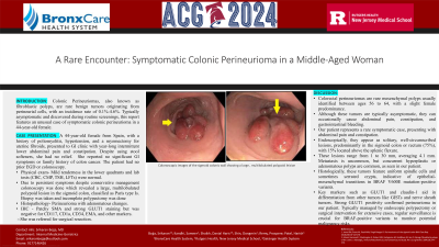
Introduction: Colonic Perineuriomas, also known as fibroblastic polyps, are rare benign tumors originating from perineurial cells, with an incidence rate of 0.1%-4.6%. Typically asymptomatic and discovered during routine screenings, this report features an unusual case of symptomatic colonic perineurioma in a 44-year-old female.
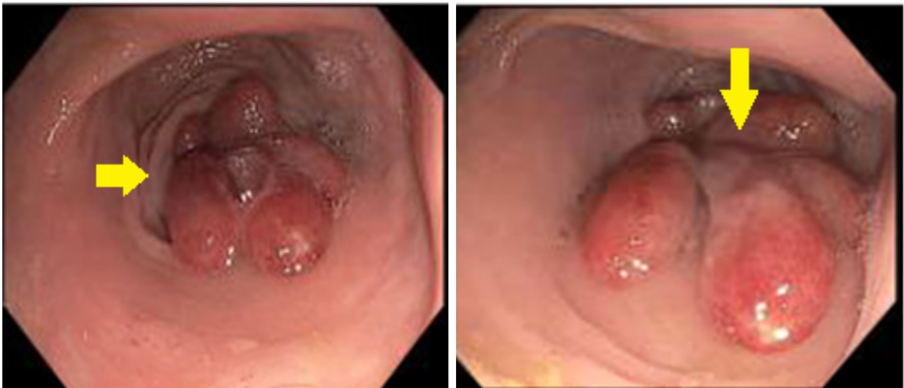
Case Description/Methods: A 44-year-old female from Spain, with a history of poliomyelitis, hypertension, and a myomectomy for uterine fibroids, presented to GI clinic with year-long intermittent lower abdominal pain and constipation. Despite using stool softeners, she had no relief. She reported no significant GI symptoms or family history of colon cancer. The patient had no prior gastroscopy or colonoscopy. Examination revealed mild tenderness in the lower quadrants and lab tests (CBC, CMP, TSH, LFTs) were normal. She was advised to increase dietary fiber and use laxatives. However, symptoms persisted, and a colonoscopy uncovered a large, multilobulated polypoid lesion in the sigmoid colon, classified as Paris type Is. A biopsy confirmed perineurioma with adenomatous changes. Immunohistochemical analysis showed patchy SMA and strong GLUT1 staining but was negative for CD117, CD1a, CD34, EMA, and other markers. She was referred for surgical resection.
Discussion: Colorectal perineuriomas are rare mesenchymal polyps usually identified between ages 56 to 64, with a slight female predominance. Although these tumors are typically asymptomatic, they can occasionally cause abdominal pain, constipation, and gastrointestinal bleeding. Our patient represents a rare symptomatic case, presenting with abdominal pain and constipation. Endoscopically, they appear as solitary, well-circumscribed lesions, predominantly in the sigmoid colon or rectum (75%), with 15% located above the splenic flexure. These lesions range from 1 to 30 mm, averaging 4.1 mm. Metastasis is uncommon, but concurrent hyperplastic or adenomatous polyps are common, as seen in our patient. Histologically, these tumors feature uniform spindle cells and sometimes serrated crypts, indicative of epithelial-mesenchymal transitions in BRAF V600E mutation-positive variants. Key markers such as GLUT1 and claudin-1 aid in differentiation from other tumors like GISTs and nerve sheath tumors. Strong GLUT1 positivity confirmed perineurioma in our patient. Typically managed by endoscopic polypectomy or surgical intervention for extensive cases, regular surveillance is crucial for BRAF-positive variants to monitor potential malignancy risks.

Disclosures:
Srikaran Bojja, MD1, Sameer Kandhi, MD1, Danial Haris. Shaikh, MD2, Dongmin Shin, MD1, Prospere Remy, MD3, Harish Patel, MD1. P2020 - A Rare Encounter: Symptomatic Colonic Perineurioma in a Middle-Aged Woman, ACG 2024 Annual Scientific Meeting Abstracts. Philadelphia, PA: American College of Gastroenterology.
1BronxCare Health System, Bronx, NY; 2Mayo Clinic, Cave Creek, AZ; 3Bronx Lebanon Hospital Center, New York, NY
Introduction: Colonic Perineuriomas, also known as fibroblastic polyps, are rare benign tumors originating from perineurial cells, with an incidence rate of 0.1%-4.6%. Typically asymptomatic and discovered during routine screenings, this report features an unusual case of symptomatic colonic perineurioma in a 44-year-old female.
Case Description/Methods: A 44-year-old female from Spain, with a history of poliomyelitis, hypertension, and a myomectomy for uterine fibroids, presented to GI clinic with year-long intermittent lower abdominal pain and constipation. Despite using stool softeners, she had no relief. She reported no significant GI symptoms or family history of colon cancer. The patient had no prior gastroscopy or colonoscopy. Examination revealed mild tenderness in the lower quadrants and lab tests (CBC, CMP, TSH, LFTs) were normal. She was advised to increase dietary fiber and use laxatives. However, symptoms persisted, and a colonoscopy uncovered a large, multilobulated polypoid lesion in the sigmoid colon, classified as Paris type Is. A biopsy confirmed perineurioma with adenomatous changes. Immunohistochemical analysis showed patchy SMA and strong GLUT1 staining but was negative for CD117, CD1a, CD34, EMA, and other markers. She was referred for surgical resection.
Discussion: Colorectal perineuriomas are rare mesenchymal polyps usually identified between ages 56 to 64, with a slight female predominance. Although these tumors are typically asymptomatic, they can occasionally cause abdominal pain, constipation, and gastrointestinal bleeding. Our patient represents a rare symptomatic case, presenting with abdominal pain and constipation. Endoscopically, they appear as solitary, well-circumscribed lesions, predominantly in the sigmoid colon or rectum (75%), with 15% located above the splenic flexure. These lesions range from 1 to 30 mm, averaging 4.1 mm. Metastasis is uncommon, but concurrent hyperplastic or adenomatous polyps are common, as seen in our patient. Histologically, these tumors feature uniform spindle cells and sometimes serrated crypts, indicative of epithelial-mesenchymal transitions in BRAF V600E mutation-positive variants. Key markers such as GLUT1 and claudin-1 aid in differentiation from other tumors like GISTs and nerve sheath tumors. Strong GLUT1 positivity confirmed perineurioma in our patient. Typically managed by endoscopic polypectomy or surgical intervention for extensive cases, regular surveillance is crucial for BRAF-positive variants to monitor potential malignancy risks.

Figure: Colonoscopic image showing a large, multilobulated polypoid leision in sigmoid colon
Disclosures:
Srikaran Bojja indicated no relevant financial relationships.
Sameer Kandhi indicated no relevant financial relationships.
Danial Shaikh indicated no relevant financial relationships.
Dongmin Shin indicated no relevant financial relationships.
Prospere Remy indicated no relevant financial relationships.
Harish Patel indicated no relevant financial relationships.
Srikaran Bojja, MD1, Sameer Kandhi, MD1, Danial Haris. Shaikh, MD2, Dongmin Shin, MD1, Prospere Remy, MD3, Harish Patel, MD1. P2020 - A Rare Encounter: Symptomatic Colonic Perineurioma in a Middle-Aged Woman, ACG 2024 Annual Scientific Meeting Abstracts. Philadelphia, PA: American College of Gastroenterology.

