Monday Poster Session
Category: Colon
P1954 - Wall Thickening of the Lower Gastrointestinal Tract on CT Imaging: What to Expect on Colonoscopy?
Monday, October 28, 2024
10:30 AM - 4:00 PM ET
Location: Exhibit Hall E

Has Audio

Osama Alshakhatreh, MD
Albany Medical Center
Albany, NY
Presenting Author(s)
Osama Alshakhatreh, MD1, Khaled Elsokary, DO1, Ibrahim Mohammed, MD1, Shunsa Tarar, DO1, Tyler Richter, MS1, Paul J. Feustel, PhD1, Madeline Cleary, DO1, Stephen Hasak, MD, MPH2
1Albany Medical Center, Albany, NY; 2Albany Medical College, Albany, NY
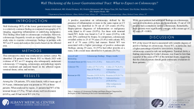
Introduction: Wall thickening (WT) of the lower gastrointestinal (GI) tract is a relatively common finding on computed tomography (CT) imaging, suggesting inflammation or underlying malignancy. This finding often leads to colonoscopic evaluation. However, colonoscopy frequently reveals no significant pathology. This study aims to examine colonoscopy findings in patients with WT on CT scans and analyze the results based on the affected area.
Methods: This retrospective study was conducted at a tertiary center. We identified 120 patients from March 2021 to March 2023 with evidence of WT on CT imaging who subsequently underwent colonoscopy. CT imaging, colonoscopy, and pathology reports were examined and analyzed based on the affected region: terminal ileum, colon, and rectum.
Results: Among the 120 patients, 55% were female, with a mean age of 55.9 years. Abdominal pain was present in 75% of these patients. When analyzed by region, 21 patients had WT of the terminal ileum (17.5%), 79 had colonic wall involvement (65.8%), and 29 had rectal WT (24.1%).
A positive association on colonoscopy, defined by the presence of inflammation or mass in the same region as CT imaging, was identified in 54 out of 129 events (41.8%). Biopsy-proven diagnoses of inflammation or malignancy were found in 45 cases (34.9%). For those with terminal ileum WT, ileitis was found in 5 of 21 cases (23.8%), with only 19% confirmed by biopsy. In comparison, colonoscopy revealed colitis in 25 of 79 cases (31.6%) with colonic WT, and 8 of 79 cases (10%) had masses. Rectal WT was associated with a higher percentage of positive endoscopic findings; among 29 cases, 16 (55%) had either proctitis or a mass, with a rectal mass identified in 6 patients (20.7%).
While most patients had additional findings on colonoscopy, such as diverticulosis, polyps, and hemorrhoids, 15 out of 120 patients had a completely normal colonoscopy. Remarkably, these patients were significantly younger, with a mean age of 42 years (P = 0.002).
Detailed findings are summarized in Table 1.
Discussion: WT of the lower GI tract is associated with variable degrees of positive findings on colonoscopy. Rectal WT, in particular, had a higher percentage of positive associations, deeming colonoscopy crucial to rule out malignancy. Terminal ileitis, a finding concerning for Crohn’s disease, was found in less than a quarter of the cases of WT of the terminal ileum, suggesting that the clinical picture should guide endoscopic evaluation in these patients.
Note: The table for this abstract can be viewed in the ePoster Gallery section of the ACG 2024 ePoster Site or in The American Journal of Gastroenterology's abstract supplement issue, both of which will be available starting October 27, 2024.
Disclosures:
Osama Alshakhatreh, MD1, Khaled Elsokary, DO1, Ibrahim Mohammed, MD1, Shunsa Tarar, DO1, Tyler Richter, MS1, Paul J. Feustel, PhD1, Madeline Cleary, DO1, Stephen Hasak, MD, MPH2. P1954 - Wall Thickening of the Lower Gastrointestinal Tract on CT Imaging: What to Expect on Colonoscopy?, ACG 2024 Annual Scientific Meeting Abstracts. Philadelphia, PA: American College of Gastroenterology.
1Albany Medical Center, Albany, NY; 2Albany Medical College, Albany, NY
Introduction: Wall thickening (WT) of the lower gastrointestinal (GI) tract is a relatively common finding on computed tomography (CT) imaging, suggesting inflammation or underlying malignancy. This finding often leads to colonoscopic evaluation. However, colonoscopy frequently reveals no significant pathology. This study aims to examine colonoscopy findings in patients with WT on CT scans and analyze the results based on the affected area.
Methods: This retrospective study was conducted at a tertiary center. We identified 120 patients from March 2021 to March 2023 with evidence of WT on CT imaging who subsequently underwent colonoscopy. CT imaging, colonoscopy, and pathology reports were examined and analyzed based on the affected region: terminal ileum, colon, and rectum.
Results: Among the 120 patients, 55% were female, with a mean age of 55.9 years. Abdominal pain was present in 75% of these patients. When analyzed by region, 21 patients had WT of the terminal ileum (17.5%), 79 had colonic wall involvement (65.8%), and 29 had rectal WT (24.1%).
A positive association on colonoscopy, defined by the presence of inflammation or mass in the same region as CT imaging, was identified in 54 out of 129 events (41.8%). Biopsy-proven diagnoses of inflammation or malignancy were found in 45 cases (34.9%). For those with terminal ileum WT, ileitis was found in 5 of 21 cases (23.8%), with only 19% confirmed by biopsy. In comparison, colonoscopy revealed colitis in 25 of 79 cases (31.6%) with colonic WT, and 8 of 79 cases (10%) had masses. Rectal WT was associated with a higher percentage of positive endoscopic findings; among 29 cases, 16 (55%) had either proctitis or a mass, with a rectal mass identified in 6 patients (20.7%).
While most patients had additional findings on colonoscopy, such as diverticulosis, polyps, and hemorrhoids, 15 out of 120 patients had a completely normal colonoscopy. Remarkably, these patients were significantly younger, with a mean age of 42 years (P = 0.002).
Detailed findings are summarized in Table 1.
Discussion: WT of the lower GI tract is associated with variable degrees of positive findings on colonoscopy. Rectal WT, in particular, had a higher percentage of positive associations, deeming colonoscopy crucial to rule out malignancy. Terminal ileitis, a finding concerning for Crohn’s disease, was found in less than a quarter of the cases of WT of the terminal ileum, suggesting that the clinical picture should guide endoscopic evaluation in these patients.
Note: The table for this abstract can be viewed in the ePoster Gallery section of the ACG 2024 ePoster Site or in The American Journal of Gastroenterology's abstract supplement issue, both of which will be available starting October 27, 2024.
Disclosures:
Osama Alshakhatreh indicated no relevant financial relationships.
Khaled Elsokary indicated no relevant financial relationships.
Ibrahim Mohammed indicated no relevant financial relationships.
Shunsa Tarar indicated no relevant financial relationships.
Tyler Richter indicated no relevant financial relationships.
Paul Feustel indicated no relevant financial relationships.
Madeline Cleary indicated no relevant financial relationships.
Stephen Hasak indicated no relevant financial relationships.
Osama Alshakhatreh, MD1, Khaled Elsokary, DO1, Ibrahim Mohammed, MD1, Shunsa Tarar, DO1, Tyler Richter, MS1, Paul J. Feustel, PhD1, Madeline Cleary, DO1, Stephen Hasak, MD, MPH2. P1954 - Wall Thickening of the Lower Gastrointestinal Tract on CT Imaging: What to Expect on Colonoscopy?, ACG 2024 Annual Scientific Meeting Abstracts. Philadelphia, PA: American College of Gastroenterology.
