Monday Poster Session
Category: Biliary/Pancreas
P1787 - Lost and Found: A Dropped Gallstone-Induced Perihepatic Abscess Post-Cholecystectomy
Monday, October 28, 2024
10:30 AM - 4:00 PM ET
Location: Exhibit Hall E


Razan Aburumman, MD
Mayo Clinic
Rochester, MN
Presenting Author(s)
Razan Aburumman, MD1, Karim Al Annan, MD2, Rudy Mrad, MD1, Khushboo Gala, MBBS3
1Mayo Clinic, Rochester, MN; 2Mayo Clinic, Hartford, CT; 3Mayo Clinic School of Graduate Medical Education, Rochester, MN
Introduction: Gallstone spillage is a recognized complication that occurs in up to 40% of laparoscopic cholecystectomy procedures and can lead to various complications. We describe a rare case of a perihepatic abscess resulting from an unretrieved gallstone, 14 months post-surgery.
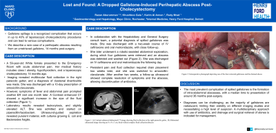
Case Description/Methods: A 59-year-old White female presented to the Emergency Room with acute abdominal pain. Her medical history includes class I obesity, hypothyroidism, and a laparoscopic cholecystectomy 14 months ago. She had no significant travel or substance use history. Imaging revealed multilocular fluid collection in the right paracolic gutter, and a diagnosis of duodenal diverticulitis was made. She was discharged with a 10-day prescription of amoxicillin-clavulanate. However, symptoms of fever and abdominal pain prompted another ER visit one month later. A Contrast-enhanced CT showed a significant increase in the size of the fluid collection (Figure 1A). [1] Laboratory results revealed leukocytosis and slightly elevated ALT. She was admitted and started on piperacillin/tazobactam. Ultrasound-guided aspiration revealed purulent material, with cultures growing E. coli and Bacteroides fragilis. The cause of the hepatic abscess was unclear, with no sources detected by CT or ultrasound. In collaboration with the Hepatobiliary and General Surgery consult team, a potential diagnosis of spilled gallstones was made. She was discharged with a two-week course of IV ceftriaxone and oral metronidazole, with close follow-up. She later underwent a robotic-assisted abdominal exploration, during which four gallstones were retrieved and an abscess was debrided and washed out (Figure 1B). She was discharged on IV ceftriaxone and oral metronidazole the following day. Persistent pain and fluid collection required drain placement two weeks later, and she was switched to amoxicillin-clavulanate. After another two weeks, a follow-up ultrasound showed complete resolution of symptoms and the abscess, allowing discontinuation of antibiotics.
Discussion: The most prevalent complication of spilled gallstones is the formation of intra-abdominal abscesses, with a median time to presentation of around 36 months post-surgery. Diagnoses can be challenging; as the majority of gallstones are radiolucent, limiting their visibility on different imaging studies and necessitating a high level of suspicion. A multidisciplinary approach with the use of antibiotics, and drainage and surgical retrieval of stones is indicated for management.

Disclosures:
Razan Aburumman, MD1, Karim Al Annan, MD2, Rudy Mrad, MD1, Khushboo Gala, MBBS3. P1787 - Lost and Found: A Dropped Gallstone-Induced Perihepatic Abscess Post-Cholecystectomy, ACG 2024 Annual Scientific Meeting Abstracts. Philadelphia, PA: American College of Gastroenterology.
1Mayo Clinic, Rochester, MN; 2Mayo Clinic, Hartford, CT; 3Mayo Clinic School of Graduate Medical Education, Rochester, MN
Introduction: Gallstone spillage is a recognized complication that occurs in up to 40% of laparoscopic cholecystectomy procedures and can lead to various complications. We describe a rare case of a perihepatic abscess resulting from an unretrieved gallstone, 14 months post-surgery.
Case Description/Methods: A 59-year-old White female presented to the Emergency Room with acute abdominal pain. Her medical history includes class I obesity, hypothyroidism, and a laparoscopic cholecystectomy 14 months ago. She had no significant travel or substance use history. Imaging revealed multilocular fluid collection in the right paracolic gutter, and a diagnosis of duodenal diverticulitis was made. She was discharged with a 10-day prescription of amoxicillin-clavulanate. However, symptoms of fever and abdominal pain prompted another ER visit one month later. A Contrast-enhanced CT showed a significant increase in the size of the fluid collection (Figure 1A). [1] Laboratory results revealed leukocytosis and slightly elevated ALT. She was admitted and started on piperacillin/tazobactam. Ultrasound-guided aspiration revealed purulent material, with cultures growing E. coli and Bacteroides fragilis. The cause of the hepatic abscess was unclear, with no sources detected by CT or ultrasound. In collaboration with the Hepatobiliary and General Surgery consult team, a potential diagnosis of spilled gallstones was made. She was discharged with a two-week course of IV ceftriaxone and oral metronidazole, with close follow-up. She later underwent a robotic-assisted abdominal exploration, during which four gallstones were retrieved and an abscess was debrided and washed out (Figure 1B). She was discharged on IV ceftriaxone and oral metronidazole the following day. Persistent pain and fluid collection required drain placement two weeks later, and she was switched to amoxicillin-clavulanate. After another two weeks, a follow-up ultrasound showed complete resolution of symptoms and the abscess, allowing discontinuation of antibiotics.
Discussion: The most prevalent complication of spilled gallstones is the formation of intra-abdominal abscesses, with a median time to presentation of around 36 months post-surgery. Diagnoses can be challenging; as the majority of gallstones are radiolucent, limiting their visibility on different imaging studies and necessitating a high level of suspicion. A multidisciplinary approach with the use of antibiotics, and drainage and surgical retrieval of stones is indicated for management.

Figure: Figure 1: A) Contrast- enhanced abdominal CT image demonstrating multilocular fluid collection in the right paracolic gutter (arrow). B) Robotic-assisted abdominal exploration; B1) Purulent fluid being suctioned (arrow); B2) One of the four stones retrieved (circle).
Disclosures:
Razan Aburumman indicated no relevant financial relationships.
Karim Al Annan indicated no relevant financial relationships.
Rudy Mrad indicated no relevant financial relationships.
Khushboo Gala indicated no relevant financial relationships.
Razan Aburumman, MD1, Karim Al Annan, MD2, Rudy Mrad, MD1, Khushboo Gala, MBBS3. P1787 - Lost and Found: A Dropped Gallstone-Induced Perihepatic Abscess Post-Cholecystectomy, ACG 2024 Annual Scientific Meeting Abstracts. Philadelphia, PA: American College of Gastroenterology.
