Monday Poster Session
Category: Biliary/Pancreas
P1790 - A Rare Case of a Rapidly Expanding High-Grade Gallbladder Neuroendocrine Tumor
Monday, October 28, 2024
10:30 AM - 4:00 PM ET
Location: Exhibit Hall E

Has Audio
- SD
Saurabh Dharmadhikari, DO
Thomas Jefferson Health
Newark, DE
Presenting Author(s)
Saurabh Dharmadhikari, DO1, Collin Reynolds, DO2, Edward Bley, DO3, Richard Walters, DO4, Jihui Qiu, MD5
1Thomas Jefferson Health, Newark, DE; 2Thomas Jefferson Health, Stratford, NJ; 3Jefferson Health, Washington Township, NJ; 4Thomas Jefferson Health, Voorhees, NJ; 5Thomas Jefferson Health, Cherry Hill, NJ
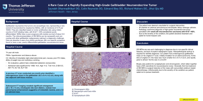
Introduction: Gallbladder neuroendocrine tumors are exceedingly rare, representing a rare subset of neuroendocrine neoplasms (NENs), with an incidence of 0.74 per 10000. They are classified based on tumor proliferative rate using mitotic counts or Ki-67 labeling index, with Ki-67 > 30% considered poorly differentiated. NENs have a poor prognosis with median survival of about 9.8 months. The clinical presentation of GB NENs can be nonspecific and can overlap with other gallbladder malignancies making it a diagnostic challenge. Here we describe a case of an elderly gentleman with non-specific symptoms found to have a rare, poorly differentiated gallbladder neuroendocrine tumor.
Case Description/Methods: A 74 year old male with hypertension and tobacco abuse presented from home with a few months of intractable right sided abdominal pain, nausea, poor PO intake, 40lbs of weight loss and nonbilious vomiting. On evaluation, he had a distended abdomen and jaundice. Admission labs were significant for WBC 19.5, Hgb 11.6, T bili 10.6, D Bili 6.5, AST 74, ALT 36, ALP 729. A previous CT scan conducted one month prior identified a heterogeneous mass in the gallbladder (8.5 x 9.4 x 4.2 cm), along with two adjacent soft tissue masses. Subsequent CT imaging revealed significant enlargement (22 x 19 x 13 cm), intrahepatic bile duct dilation, nodular liver parenchymal enhancement suggestive of metastatic lesions, and a 1.6 cm adrenal mass. The patient was deemed unsuitable for surgical intervention. IR was consulted for a biopsy and insertion of an external percutaneous transhepatic biliary drainage catheter. Pathology confirmed diagnosis of high-grade neuroendocrine tumor consistent with small cell neuroendocrine carcinoma with Ki-67 >80%. Due to the severity of his condition, the patient declined treatment and opted for home hospice care.
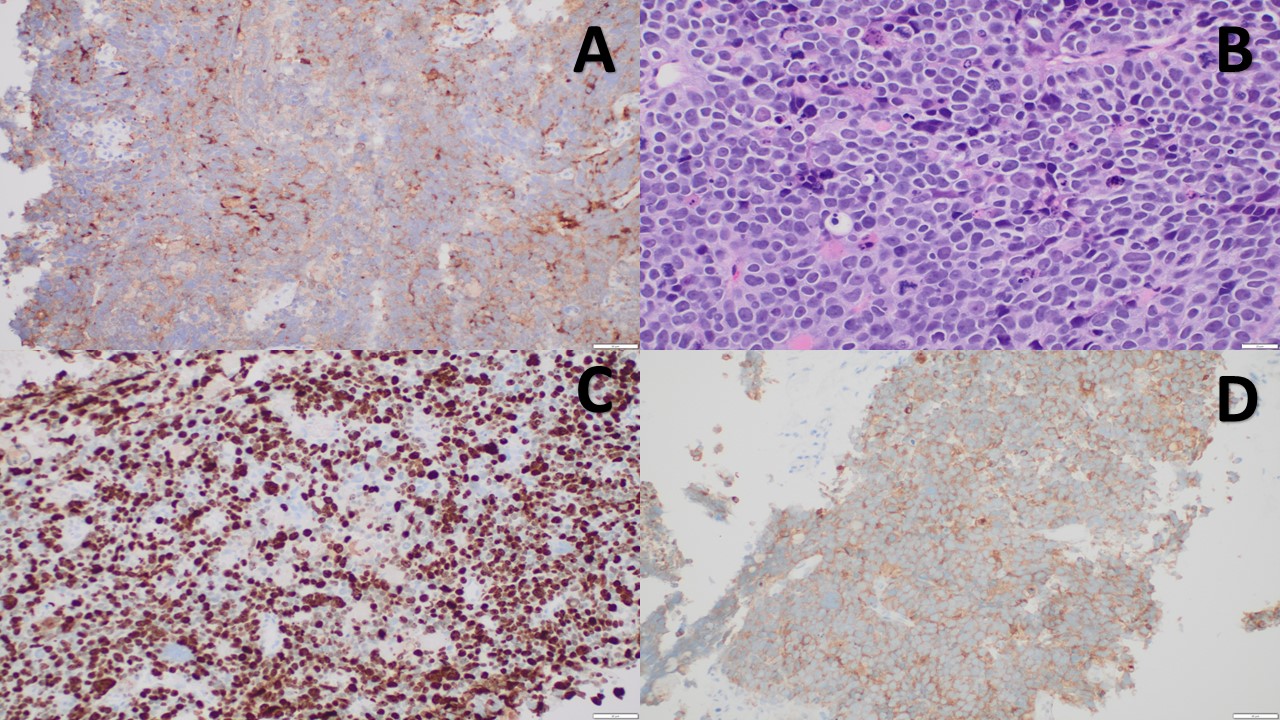
Discussion: GB NENs are rare and challenging to diagnose due to non-specific clinical features, and lack of explicit radiographic signs. Histopathological workup is required for definite diagnosis. Our patient had a heterogenous gallbladder mass on his original CT scan, initially mistaken for an adenocarcinoma. Biopsy was positive for synaptophysin and chromogranin, which taken together have a 95% sensitivity for neuroendocrine tumor. Generally first line treatment of GB NEN is surgical resection of the mass with concomitant platinum based chemotherapy. More research is needed to improve early diagnosis and streamline treatment guidelines for gallbladder neuroendocrine carcinoma.

Disclosures:
Saurabh Dharmadhikari, DO1, Collin Reynolds, DO2, Edward Bley, DO3, Richard Walters, DO4, Jihui Qiu, MD5. P1790 - A Rare Case of a Rapidly Expanding High-Grade Gallbladder Neuroendocrine Tumor, ACG 2024 Annual Scientific Meeting Abstracts. Philadelphia, PA: American College of Gastroenterology.
1Thomas Jefferson Health, Newark, DE; 2Thomas Jefferson Health, Stratford, NJ; 3Jefferson Health, Washington Township, NJ; 4Thomas Jefferson Health, Voorhees, NJ; 5Thomas Jefferson Health, Cherry Hill, NJ
Introduction: Gallbladder neuroendocrine tumors are exceedingly rare, representing a rare subset of neuroendocrine neoplasms (NENs), with an incidence of 0.74 per 10000. They are classified based on tumor proliferative rate using mitotic counts or Ki-67 labeling index, with Ki-67 > 30% considered poorly differentiated. NENs have a poor prognosis with median survival of about 9.8 months. The clinical presentation of GB NENs can be nonspecific and can overlap with other gallbladder malignancies making it a diagnostic challenge. Here we describe a case of an elderly gentleman with non-specific symptoms found to have a rare, poorly differentiated gallbladder neuroendocrine tumor.
Case Description/Methods: A 74 year old male with hypertension and tobacco abuse presented from home with a few months of intractable right sided abdominal pain, nausea, poor PO intake, 40lbs of weight loss and nonbilious vomiting. On evaluation, he had a distended abdomen and jaundice. Admission labs were significant for WBC 19.5, Hgb 11.6, T bili 10.6, D Bili 6.5, AST 74, ALT 36, ALP 729. A previous CT scan conducted one month prior identified a heterogeneous mass in the gallbladder (8.5 x 9.4 x 4.2 cm), along with two adjacent soft tissue masses. Subsequent CT imaging revealed significant enlargement (22 x 19 x 13 cm), intrahepatic bile duct dilation, nodular liver parenchymal enhancement suggestive of metastatic lesions, and a 1.6 cm adrenal mass. The patient was deemed unsuitable for surgical intervention. IR was consulted for a biopsy and insertion of an external percutaneous transhepatic biliary drainage catheter. Pathology confirmed diagnosis of high-grade neuroendocrine tumor consistent with small cell neuroendocrine carcinoma with Ki-67 >80%. Due to the severity of his condition, the patient declined treatment and opted for home hospice care.
Discussion: GB NENs are rare and challenging to diagnose due to non-specific clinical features, and lack of explicit radiographic signs. Histopathological workup is required for definite diagnosis. Our patient had a heterogenous gallbladder mass on his original CT scan, initially mistaken for an adenocarcinoma. Biopsy was positive for synaptophysin and chromogranin, which taken together have a 95% sensitivity for neuroendocrine tumor. Generally first line treatment of GB NEN is surgical resection of the mass with concomitant platinum based chemotherapy. More research is needed to improve early diagnosis and streamline treatment guidelines for gallbladder neuroendocrine carcinoma.

Figure: A) Chromogranin 200x
B) Hematoxylin and Eosin 400x
C) Ki-67
D) Synaptophysin 200x
B) Hematoxylin and Eosin 400x
C) Ki-67
D) Synaptophysin 200x
Disclosures:
Saurabh Dharmadhikari indicated no relevant financial relationships.
Collin Reynolds indicated no relevant financial relationships.
Edward Bley indicated no relevant financial relationships.
Richard Walters indicated no relevant financial relationships.
Jihui Qiu indicated no relevant financial relationships.
Saurabh Dharmadhikari, DO1, Collin Reynolds, DO2, Edward Bley, DO3, Richard Walters, DO4, Jihui Qiu, MD5. P1790 - A Rare Case of a Rapidly Expanding High-Grade Gallbladder Neuroendocrine Tumor, ACG 2024 Annual Scientific Meeting Abstracts. Philadelphia, PA: American College of Gastroenterology.
