Monday Poster Session
Category: Biliary/Pancreas
P1795 - Pancreatic Panniculitis: An Unusual Initial Presentation of Intraductal Papillary Mucinous Neoplasm (IPMN)
Monday, October 28, 2024
10:30 AM - 4:00 PM ET
Location: Exhibit Hall E

Has Audio

Mohamad Hijazi, MD
TriHealth Good Samaritan Hospital
West Chester, OH
Presenting Author(s)
Mohamad Hijazi, MD1, M Kenan Rahima, MD1, Mhd Kutaiba Albuni, MD2, Tarek Aboursheid, MD3, Venkata Muddana, MD4
1TriHealth Good Samaritan Hospital, Cincinnati, OH; 2TriHealth, Doha, Ad Dawhah, Qatar; 3Ascension Saint Francis Hospital, Evanston, IL; 4TriHealth, Cincinnati, OH
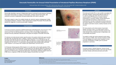
Introduction: Panniculitis describes a group of inflammatory lesions involving the subcutaneous fat tissue. Pancreatic panniculitis, a subtype of panniculitis with adipocyte necrosis, was initially misdiagnosed as erythema nodosum in an elderly female. Further assessment led to the diagnosis of intraductal papillary mucinous neoplasm (IPMN).
Case Description/Methods: An 80-year-old female presented with bilateral painful lower extremity lesions initially diagnosed as erythema nodosum (EN) and treated with steroids, which partially improved her symptoms. Upon worsening of the lesions and development of ulceration after stopping steroids, she was treated with Dapsone for resistant EN. A dermatologist's assessment and subsequent skin biopsy revealed pancreatic panniculitis, characterized by diffuse lipocyte degeneration and early calcification, indicative of a ghost cell change. Further evaluation via MRI and endoscopic ultrasonography (EUS) of the abdomen identified a 3 cm cystic lesion with septation in the pancreatic neck, with a diffusely dilated pancreatic duct, suggesting a mixed duct intraductal papillary mucinous neoplasm (IPMN). Fine needle aspiration (FNA) cytology was negative for malignant cells, but the cyst fluid's CEA level was high at 528. Based on the EUS findings and elevated CEA level, a diagnosis of IPMN was established.
Discussion: Pancreatic panniculitis (PP) is a rare subtype of panniculitis associated with pancreatic diseases, typically affecting males and often linked to alcoholism. PP usually presents as multiple erythematous or red-brown nodules on the lower extremities and buttocks, characterized by adipocyte necrosis and calcium deposits. It rarely appears as the initial symptom of pancreatic illness, commonly associating with acute or chronic pancreatitis, which was not evident in our patient. Instead, PP led to the diagnosis of intraductal papillary mucinous neoplasm (IPMN). This patient’s skin lesions appeared months before any IPMN symptoms, with painless jaundice emerging later. The initial CT scan missed the neoplasm, which was later detected by MRI. Physicians should recognize extrapancreatic manifestations of IPMN for early detection. Detailed physical examinations remain crucial despite advanced diagnostic tools.

Disclosures:
Mohamad Hijazi, MD1, M Kenan Rahima, MD1, Mhd Kutaiba Albuni, MD2, Tarek Aboursheid, MD3, Venkata Muddana, MD4. P1795 - Pancreatic Panniculitis: An Unusual Initial Presentation of Intraductal Papillary Mucinous Neoplasm (IPMN), ACG 2024 Annual Scientific Meeting Abstracts. Philadelphia, PA: American College of Gastroenterology.
1TriHealth Good Samaritan Hospital, Cincinnati, OH; 2TriHealth, Doha, Ad Dawhah, Qatar; 3Ascension Saint Francis Hospital, Evanston, IL; 4TriHealth, Cincinnati, OH
Introduction: Panniculitis describes a group of inflammatory lesions involving the subcutaneous fat tissue. Pancreatic panniculitis, a subtype of panniculitis with adipocyte necrosis, was initially misdiagnosed as erythema nodosum in an elderly female. Further assessment led to the diagnosis of intraductal papillary mucinous neoplasm (IPMN).
Case Description/Methods: An 80-year-old female presented with bilateral painful lower extremity lesions initially diagnosed as erythema nodosum (EN) and treated with steroids, which partially improved her symptoms. Upon worsening of the lesions and development of ulceration after stopping steroids, she was treated with Dapsone for resistant EN. A dermatologist's assessment and subsequent skin biopsy revealed pancreatic panniculitis, characterized by diffuse lipocyte degeneration and early calcification, indicative of a ghost cell change. Further evaluation via MRI and endoscopic ultrasonography (EUS) of the abdomen identified a 3 cm cystic lesion with septation in the pancreatic neck, with a diffusely dilated pancreatic duct, suggesting a mixed duct intraductal papillary mucinous neoplasm (IPMN). Fine needle aspiration (FNA) cytology was negative for malignant cells, but the cyst fluid's CEA level was high at 528. Based on the EUS findings and elevated CEA level, a diagnosis of IPMN was established.
Discussion: Pancreatic panniculitis (PP) is a rare subtype of panniculitis associated with pancreatic diseases, typically affecting males and often linked to alcoholism. PP usually presents as multiple erythematous or red-brown nodules on the lower extremities and buttocks, characterized by adipocyte necrosis and calcium deposits. It rarely appears as the initial symptom of pancreatic illness, commonly associating with acute or chronic pancreatitis, which was not evident in our patient. Instead, PP led to the diagnosis of intraductal papillary mucinous neoplasm (IPMN). This patient’s skin lesions appeared months before any IPMN symptoms, with painless jaundice emerging later. The initial CT scan missed the neoplasm, which was later detected by MRI. Physicians should recognize extrapancreatic manifestations of IPMN for early detection. Detailed physical examinations remain crucial despite advanced diagnostic tools.

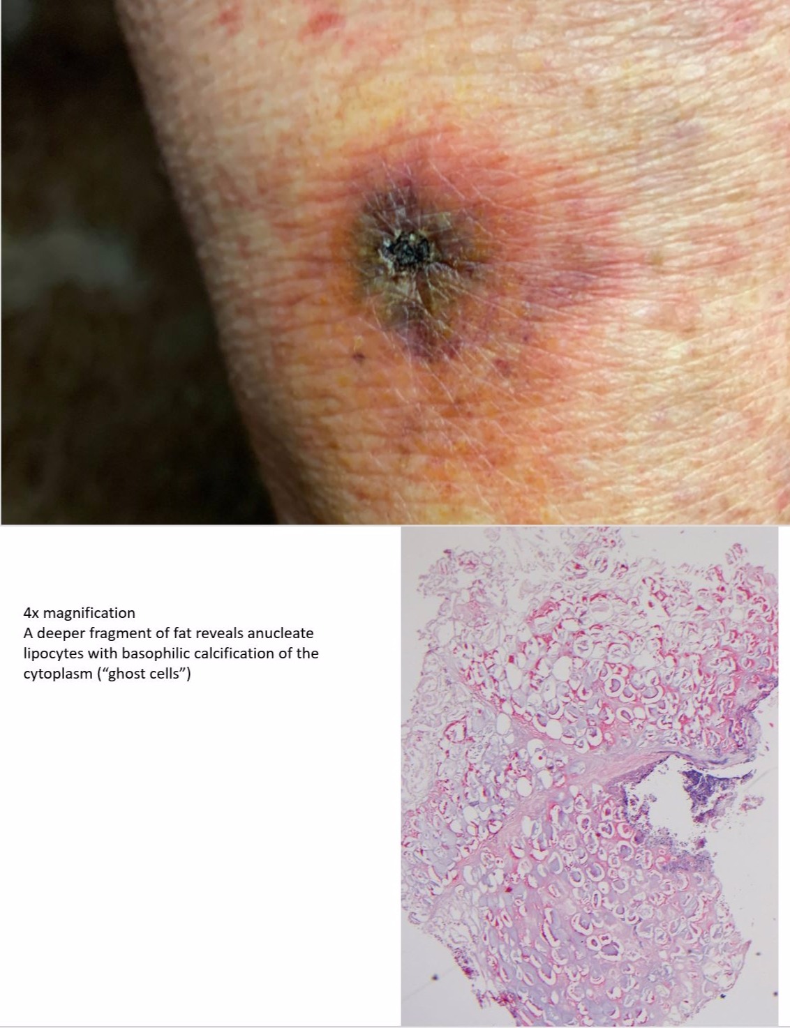
Figure: The image on the top shows the right lower extremity skin lesion after treatment with steroids
The underneeth image: 4x magnification, A deeper fragment of fat reveals anucleate lipocytes with basophilic calcification of the cytoplasm ("ghost cells")
The underneeth image: 4x magnification, A deeper fragment of fat reveals anucleate lipocytes with basophilic calcification of the cytoplasm ("ghost cells")
Disclosures:
Mohamad Hijazi indicated no relevant financial relationships.
M Kenan Rahima indicated no relevant financial relationships.
Mhd Kutaiba Albuni indicated no relevant financial relationships.
Tarek Aboursheid indicated no relevant financial relationships.
Venkata Muddana indicated no relevant financial relationships.
Mohamad Hijazi, MD1, M Kenan Rahima, MD1, Mhd Kutaiba Albuni, MD2, Tarek Aboursheid, MD3, Venkata Muddana, MD4. P1795 - Pancreatic Panniculitis: An Unusual Initial Presentation of Intraductal Papillary Mucinous Neoplasm (IPMN), ACG 2024 Annual Scientific Meeting Abstracts. Philadelphia, PA: American College of Gastroenterology.
