Sunday Poster Session
Category: Stomach
P1708 - A Case of Emphysematous Gastritis: A Deadly Disease in Diabetic Patients
Sunday, October 27, 2024
3:30 PM - 7:00 PM ET
Location: Exhibit Hall E

Has Audio

Leilah A. Tisheh, MD
Long Island Community Hospital - NYU Langone Health System
Oceanside, NY
Presenting Author(s)
Leilah A. Tisheh, MD1, Neela Qadir, MD2, Victoria Oriuwa, DO, MS2, Prakash Viswanathan, MD, FACG2, Ayse Durgun, MD2, Yuriy Takhalov, MD2
1Long Island Community Hospital - NYU Langone Health System, Oceanside, NY; 2Long Island Community Hospital - NYU Langone Health System, Patchogue, NY
Introduction: Emphysematous gastritis is a severe form of gastritis characterized by invasion of the gastric wall by gas-producing bacteria. First reported in 1889, it has been a rare phenomenon carrying a mortality rate upwards of 55% according to the literature.
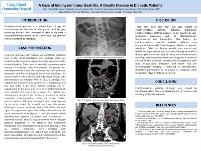
Case Description/Methods: A 66-year-old male with multiple co-morbidities including type 1 DM, atrial fibrillation, and multiple CVAs was brought into the ED for acute metabolic encephalopathy. There was no reported abdominal pain, nausea, or vomiting. Upon examination, the abdomen was soft and non-distended and his neurological exam was significant for slurred speech with a chronic left-sided facial droop. Labs demonstrated leukocytosis, a urinalysis was negative for leukocytes and positive for >1000 glucose and A1c was 8.3%. A CT brain without contrast and CT angiography of the brain and neck were performed which were negative for any acute etiology. The patient was subsequently admitted for further assessment of acute metabolic encephalopathy; ruling out stroke. Further work-up with an MRI was performed which was negative for an acute stroke. By hospital day three, the patient developed nausea, vomiting, abdominal distension with signs of progressive sepsis and diabetic ketoacidosis. An initial abdominal x-ray was performed which demonstrated gaseous distension and a follow-up CT abdomen without contrast was performed which revealed gaseous distention in the stomach and duodenum compatible with emphysematous gastritis. Empiric antibiotics were initiated with piperacillin/tazobactam, the patient was kept NPO, and fluid resuscitated. The patient rapidly decompensated due to progressive sepsis and expired shortly thereafter.
Discussion: There have been less than 100 case reports of emphysematous gastritis between 1980-2022. Emphysematous gastritis appears to be caused by gas-producing organisms such as Staphylococcus, Streptococcus, and Clostridium. Risk factors for emphysematous gastritis include diabetes and immunodeficiency which predisposes patients to systemic infections. Other risk factors include prior steroid and NSAID use, high alcohol use, and corrosive ingestion which disrupt gastric mucosa. Typical symptoms include nausea, vomiting, and abdominal distension. Diagnosis is made via a CT scan of the abdomen. Conservative management with fluid resuscitation, antibiotics, and bowel rest are recommended. Surgery is indicated in hemodynamic instability, perforation, or formation of strictures. Early recognition plays a vital role in survival.

Disclosures:
Leilah A. Tisheh, MD1, Neela Qadir, MD2, Victoria Oriuwa, DO, MS2, Prakash Viswanathan, MD, FACG2, Ayse Durgun, MD2, Yuriy Takhalov, MD2. P1708 - A Case of Emphysematous Gastritis: A Deadly Disease in Diabetic Patients, ACG 2024 Annual Scientific Meeting Abstracts. Philadelphia, PA: American College of Gastroenterology.
1Long Island Community Hospital - NYU Langone Health System, Oceanside, NY; 2Long Island Community Hospital - NYU Langone Health System, Patchogue, NY
Introduction: Emphysematous gastritis is a severe form of gastritis characterized by invasion of the gastric wall by gas-producing bacteria. First reported in 1889, it has been a rare phenomenon carrying a mortality rate upwards of 55% according to the literature.
Case Description/Methods: A 66-year-old male with multiple co-morbidities including type 1 DM, atrial fibrillation, and multiple CVAs was brought into the ED for acute metabolic encephalopathy. There was no reported abdominal pain, nausea, or vomiting. Upon examination, the abdomen was soft and non-distended and his neurological exam was significant for slurred speech with a chronic left-sided facial droop. Labs demonstrated leukocytosis, a urinalysis was negative for leukocytes and positive for >1000 glucose and A1c was 8.3%. A CT brain without contrast and CT angiography of the brain and neck were performed which were negative for any acute etiology. The patient was subsequently admitted for further assessment of acute metabolic encephalopathy; ruling out stroke. Further work-up with an MRI was performed which was negative for an acute stroke. By hospital day three, the patient developed nausea, vomiting, abdominal distension with signs of progressive sepsis and diabetic ketoacidosis. An initial abdominal x-ray was performed which demonstrated gaseous distension and a follow-up CT abdomen without contrast was performed which revealed gaseous distention in the stomach and duodenum compatible with emphysematous gastritis. Empiric antibiotics were initiated with piperacillin/tazobactam, the patient was kept NPO, and fluid resuscitated. The patient rapidly decompensated due to progressive sepsis and expired shortly thereafter.
Discussion: There have been less than 100 case reports of emphysematous gastritis between 1980-2022. Emphysematous gastritis appears to be caused by gas-producing organisms such as Staphylococcus, Streptococcus, and Clostridium. Risk factors for emphysematous gastritis include diabetes and immunodeficiency which predisposes patients to systemic infections. Other risk factors include prior steroid and NSAID use, high alcohol use, and corrosive ingestion which disrupt gastric mucosa. Typical symptoms include nausea, vomiting, and abdominal distension. Diagnosis is made via a CT scan of the abdomen. Conservative management with fluid resuscitation, antibiotics, and bowel rest are recommended. Surgery is indicated in hemodynamic instability, perforation, or formation of strictures. Early recognition plays a vital role in survival.

Figure: Abdominal x-ray (image A) demonstrating gaseous distension and CT abdomen without contrast demonstrating gaseous distention in the stomach and duodenum compatible with emphysematous gastritis (images B and C).
Disclosures:
Leilah Tisheh indicated no relevant financial relationships.
Neela Qadir indicated no relevant financial relationships.
Victoria Oriuwa indicated no relevant financial relationships.
Prakash Viswanathan indicated no relevant financial relationships.
Ayse Durgun indicated no relevant financial relationships.
Yuriy Takhalov indicated no relevant financial relationships.
Leilah A. Tisheh, MD1, Neela Qadir, MD2, Victoria Oriuwa, DO, MS2, Prakash Viswanathan, MD, FACG2, Ayse Durgun, MD2, Yuriy Takhalov, MD2. P1708 - A Case of Emphysematous Gastritis: A Deadly Disease in Diabetic Patients, ACG 2024 Annual Scientific Meeting Abstracts. Philadelphia, PA: American College of Gastroenterology.
