Sunday Poster Session
Category: Small Intestine
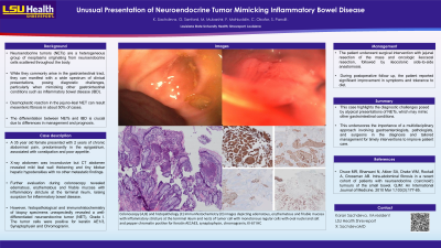
P1543 - Unusual Presentation of Neuroendocrine Tumor Mimicking Inflammatory Bowel Disease
Sunday, October 27, 2024
3:30 PM - 7:00 PM ET
Location: Exhibit Hall E

Has Audio

Karan Sachdeva, MD
LSU Health
Shreveport, LA
Presenting Author(s)
Karan Sachdeva, MD1, Gabrielle Sanford, MD2, Maryam Mubashir, MD1, Farhan Mohiuddin, MD3, Sudha Pandit, MD1
1LSU Health, Shreveport, LA; 2Louisiana State University Health, Shreveport, LA; 3LSU Health, New Orleans, LA
Introduction: Neuroendocrine tumors (NETs) are a heterogeneous group of neoplasms originating from neuroendocrine cells scattered throughout the body. While they commonly arise in the gastrointestinal tract, they can manifest with a wide spectrum of clinical presentations, posing diagnostic challenges, particularly when mimicking other gastrointestinal conditions such as inflammatory bowel disease (IBD). The differentiation between NETs and IBD is crucial due to differences in management and prognosis.
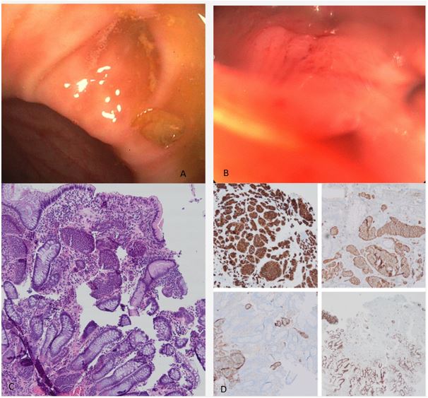
Case Description/Methods: A 35 year old female presented with 2 years of chronic abdominal pain, predominantly in the epigastrium, associated with constipation and poor appetite. X-ray abdomen was inconclusive but CT abdomen revealed mild ileal wall thickening and tiny bilobar hepatic hypodensities with no other metastatic findings. Further evaluation during colonoscopy revealed edematous, erythematous and friable mucosa with inflammatory stricture at the terminal ileum (Image A and B), raising suspicion for inflammatory bowel disease. However, histopathological and immunohistochemistry of biopsy specimens unexpectedly revealed a well-differentiated neuroendocrine tumor (NET), Grade I (Image C and D). The tumor cells were positive for keratin AE1/3, Synaptophysin and Chromogranin. The patient underwent surgical intervention with jejunal resection of the mass and oncologic ileocecal resection, followed by ileocolonic side-to-side anastomosis. Pahtology reported pT3N1 with negative margins. During postoperative period, the patient reported significant improvement in symptoms, tolerance to diet and continues to follow up.
Discussion: This case highlights the diagnostic challenges posed by atypical presentations of NETs, which may mimic other gastrointestinal conditions. This underscores the importance of a multidisciplinary approach involving gastroenterologists, pathologists, and surgeons in the diagnosis and tailored management for timely interventions to improve patient care.

Disclosures:
Karan Sachdeva, MD1, Gabrielle Sanford, MD2, Maryam Mubashir, MD1, Farhan Mohiuddin, MD3, Sudha Pandit, MD1. P1543 - Unusual Presentation of Neuroendocrine Tumor Mimicking Inflammatory Bowel Disease, ACG 2024 Annual Scientific Meeting Abstracts. Philadelphia, PA: American College of Gastroenterology.
1LSU Health, Shreveport, LA; 2Louisiana State University Health, Shreveport, LA; 3LSU Health, New Orleans, LA
Introduction: Neuroendocrine tumors (NETs) are a heterogeneous group of neoplasms originating from neuroendocrine cells scattered throughout the body. While they commonly arise in the gastrointestinal tract, they can manifest with a wide spectrum of clinical presentations, posing diagnostic challenges, particularly when mimicking other gastrointestinal conditions such as inflammatory bowel disease (IBD). The differentiation between NETs and IBD is crucial due to differences in management and prognosis.
Case Description/Methods: A 35 year old female presented with 2 years of chronic abdominal pain, predominantly in the epigastrium, associated with constipation and poor appetite. X-ray abdomen was inconclusive but CT abdomen revealed mild ileal wall thickening and tiny bilobar hepatic hypodensities with no other metastatic findings. Further evaluation during colonoscopy revealed edematous, erythematous and friable mucosa with inflammatory stricture at the terminal ileum (Image A and B), raising suspicion for inflammatory bowel disease. However, histopathological and immunohistochemistry of biopsy specimens unexpectedly revealed a well-differentiated neuroendocrine tumor (NET), Grade I (Image C and D). The tumor cells were positive for keratin AE1/3, Synaptophysin and Chromogranin. The patient underwent surgical intervention with jejunal resection of the mass and oncologic ileocecal resection, followed by ileocolonic side-to-side anastomosis. Pahtology reported pT3N1 with negative margins. During postoperative period, the patient reported significant improvement in symptoms, tolerance to diet and continues to follow up.
Discussion: This case highlights the diagnostic challenges posed by atypical presentations of NETs, which may mimic other gastrointestinal conditions. This underscores the importance of a multidisciplinary approach involving gastroenterologists, pathologists, and surgeons in the diagnosis and tailored management for timely interventions to improve patient care.

Figure: Colonoscopy (A,B) and histopathology (C) Immunhistochemistry (D) images depicting edematous, erythematous and friable mucosa with inflammatory stricture at the terminal ileum and nests of tumor cell with monotonous regular cells with oval nuclei and salt and pepper chromatin positive for Keratin AE1AE3, synaptophysin, chromogranin, KI-67 IHC
Disclosures:
Karan Sachdeva indicated no relevant financial relationships.
Gabrielle Sanford indicated no relevant financial relationships.
Maryam Mubashir indicated no relevant financial relationships.
Farhan Mohiuddin indicated no relevant financial relationships.
Sudha Pandit indicated no relevant financial relationships.
Karan Sachdeva, MD1, Gabrielle Sanford, MD2, Maryam Mubashir, MD1, Farhan Mohiuddin, MD3, Sudha Pandit, MD1. P1543 - Unusual Presentation of Neuroendocrine Tumor Mimicking Inflammatory Bowel Disease, ACG 2024 Annual Scientific Meeting Abstracts. Philadelphia, PA: American College of Gastroenterology.
