Sunday Poster Session
Category: Small Intestine
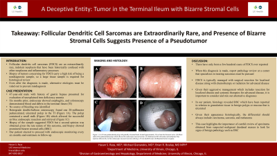
P1544 - A Deceptive Entity: Tumor in the Terminal Ileum With Bizarre Stromal Cells on Histology
Sunday, October 27, 2024
3:30 PM - 7:00 PM ET
Location: Exhibit Hall E

Has Audio
- HR
Hasan S. Raza, MD
University of Illinois
Chicago, IL
Presenting Author(s)
Hasan S. Raza, MD1, Michael Gianarakis, MD2, Brian R. Boulay, MD, MPH1
1University of Illinois, Chicago, IL; 2University of Illinois at Chicago, Chicago, IL
Introduction: Follicular dendritic cell sarcomas (FDCS) are an extraordinarily rare, indolent neoplasm that have been historically confused with other neoplasms and inflammatory processes. Biopsy of tumors concerning for FDCS carry a high risk of being a nondiagnostic sample, so a large tissue sample is required for histological review. Even after the diagnosis is made, alternative etiologies must be ruled out to prevent misdiagnosis. Herein, we present a case of an ileal mass initially diagnosed as FDCS, ultimately reclassified as a pseudotumor due to bizarre stromal cells on histopathology.
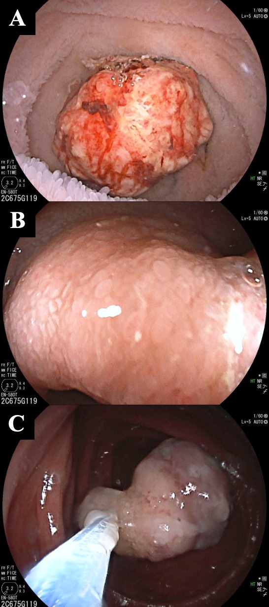
Case Description/Methods: A 47-year-old male with history of Roux-en-Y gastric bypass presented for evaluation of unexplained iron deficiency anemia. Six months prior, esophagogastroduodenoscopy (EGD) showed esophagitis, and colonoscopy demonstrated blood, erosions, and congestion in the terminal ileum (TI). No source of bleeding was identified, and a focal ulcer in the distal small bowel was found on video capsule endoscopy (VCE) three months later. Retrograde double-balloon enteroscopy found one 20-millimeter pedunculated, ulcerated polyp in the TI (Figure 1A). The polyp contained a small stalk (Figure 1B) which allowed for successful en bloc endoscopic resection and retrieval (Figure 1C). Biopsy of the sample suggested FDCS. A second opinion was obtained given the rare nature of this sarcoma, and biopsy showed a polyp with intense stromal inflammation with prominent bizarre stromal cells (BSC). Our patient elected to proceed with endoscopic monitoring every six months and continues to follow up.
Discussion: There have only been a few hundred cases of FDCS ever reported. When this diagnosis is made, expert pathology review at a center that specializes in treating sarcomas must be pursued. FDCS is typically managed with surgical resection for localized disease along with chemotherapy or radiation for advanced disease. Given their aggressive management which includes resection for localized disease and systemic therapies for advanced disease, it is important to consider and rule out alternative diagnoses. In our patient, histology revealed BSC which have been reported in relation to granulation tissue in benign polyps or mucosa that is ulcerated. Given their appearance histologically, the differential should always include carcinoma, sarcoma, and melanoma. This case highlights the importance of careful review of specimens obtained from suspected malignant duodenal masses to look for signs of benign pathology such as BSC.

Disclosures:
Hasan S. Raza, MD1, Michael Gianarakis, MD2, Brian R. Boulay, MD, MPH1. P1544 - A Deceptive Entity: Tumor in the Terminal Ileum With Bizarre Stromal Cells on Histology, ACG 2024 Annual Scientific Meeting Abstracts. Philadelphia, PA: American College of Gastroenterology.
1University of Illinois, Chicago, IL; 2University of Illinois at Chicago, Chicago, IL
Introduction: Follicular dendritic cell sarcomas (FDCS) are an extraordinarily rare, indolent neoplasm that have been historically confused with other neoplasms and inflammatory processes. Biopsy of tumors concerning for FDCS carry a high risk of being a nondiagnostic sample, so a large tissue sample is required for histological review. Even after the diagnosis is made, alternative etiologies must be ruled out to prevent misdiagnosis. Herein, we present a case of an ileal mass initially diagnosed as FDCS, ultimately reclassified as a pseudotumor due to bizarre stromal cells on histopathology.
Case Description/Methods: A 47-year-old male with history of Roux-en-Y gastric bypass presented for evaluation of unexplained iron deficiency anemia. Six months prior, esophagogastroduodenoscopy (EGD) showed esophagitis, and colonoscopy demonstrated blood, erosions, and congestion in the terminal ileum (TI). No source of bleeding was identified, and a focal ulcer in the distal small bowel was found on video capsule endoscopy (VCE) three months later. Retrograde double-balloon enteroscopy found one 20-millimeter pedunculated, ulcerated polyp in the TI (Figure 1A). The polyp contained a small stalk (Figure 1B) which allowed for successful en bloc endoscopic resection and retrieval (Figure 1C). Biopsy of the sample suggested FDCS. A second opinion was obtained given the rare nature of this sarcoma, and biopsy showed a polyp with intense stromal inflammation with prominent bizarre stromal cells (BSC). Our patient elected to proceed with endoscopic monitoring every six months and continues to follow up.
Discussion: There have only been a few hundred cases of FDCS ever reported. When this diagnosis is made, expert pathology review at a center that specializes in treating sarcomas must be pursued. FDCS is typically managed with surgical resection for localized disease along with chemotherapy or radiation for advanced disease. Given their aggressive management which includes resection for localized disease and systemic therapies for advanced disease, it is important to consider and rule out alternative diagnoses. In our patient, histology revealed BSC which have been reported in relation to granulation tissue in benign polyps or mucosa that is ulcerated. Given their appearance histologically, the differential should always include carcinoma, sarcoma, and melanoma. This case highlights the importance of careful review of specimens obtained from suspected malignant duodenal masses to look for signs of benign pathology such as BSC.

Figure: (A) 20 mm pedunculated polyp with partially ulcerated head, located approximately 10 cm from the ileocecal valve. (B) Short stalk of the pedunculated polyp which allowed for endoscopic mucosal section. (C) Submucosal lift with saline and methylene blue injection followed by en bloc endoscopic mucosal resection utilizing a 25 mm duckbill snare.
Disclosures:
Hasan Raza indicated no relevant financial relationships.
Michael Gianarakis indicated no relevant financial relationships.
Brian Boulay indicated no relevant financial relationships.
Hasan S. Raza, MD1, Michael Gianarakis, MD2, Brian R. Boulay, MD, MPH1. P1544 - A Deceptive Entity: Tumor in the Terminal Ileum With Bizarre Stromal Cells on Histology, ACG 2024 Annual Scientific Meeting Abstracts. Philadelphia, PA: American College of Gastroenterology.
