Sunday Poster Session
Category: Liver
P1285 - A Challenging Diagnosis of Hepatosplenic T-cell Lymphoma
Sunday, October 27, 2024
3:30 PM - 7:00 PM ET
Location: Exhibit Hall E

Has Audio

Bianca Thakkar, DO
University of Connecticut Health Center
Farmington, CT
Presenting Author(s)
Bianca Thakkar, DO1, Teresa Da Cunha, MD1, Danzhu Zhao, DO1, Anum Dileep, MBBS2, Marianna Scranton, DO2
1University of Connecticut Health Center, Farmington, CT; 2Hartford Hospital, Hartford, CT
Introduction: Hepatosplenic T-cell lymphoma (HSTCL) is a rare, aggressive extra nodal and systemic tumor originating from cytotoxic T cells, which is caused by the proliferation of mature T-cells that infiltrate the sinusoids of the liver and spleen. Here we present a rare case and challenging diagnosis of HSTCL in a patient with persistent jaundice.
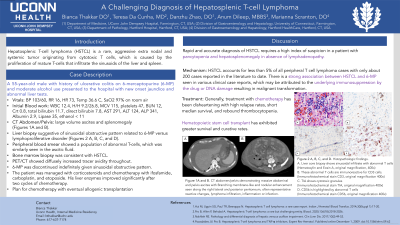
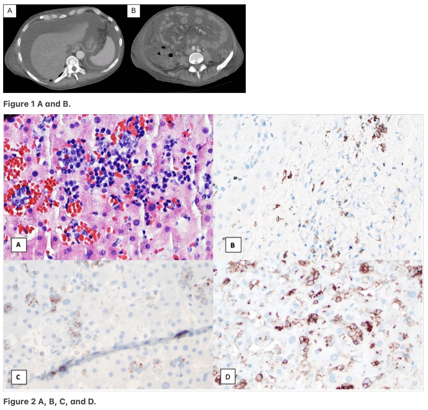
Case Description/Methods: A 55-year-old male with history of ulcerative colitis on 6-mercaptopurine (6-MP) and moderate alcohol use presented to the hospital with a few weeks of jaundice and elevated liver enzymes. The patient was previously admitted for similar symptoms and was treated for alcoholic hepatitis with steroids, which he did not respond to. During this subsequent admission, CT of the abdomen and pelvis showed large volume ascites and splenomegaly and blood work showed leukocytosis, anemia, and thrombocytopenia (Figures 1A and B). Workup for hemolytic anemia was negative. He underwent liver biopsy suggestive of sinusoidal obstructive pattern related to 6-MP versus lymphoproliferative disorder (Figures 2 A, B, C, and D). His hospital course was complicated by anemia requiring blood transfusions and fever of unclear origin. Peripheral blood smear showed a population of abnormal T-cells, which was similarly seen in the ascitic fluid. Bone marrow biopsy was consistent with HSTCL. PET/CT showed diffusely increased tracer avidity throughout. The patient was managed with multiple therapeutic paracentesis, steroids, dexamethasone, and chemotherapy with ifosfamide, carboplatin, and etoposide. He was given allopurinol for prophylaxis of tumor lysis syndrome and monitored. His liver enzymes improved significantly after two cycles of chemotherapy.
Discussion: Rapid and accurate diagnosis of HSTCL requires a high index of suspicion in a patient with pancytopenia and hepatosplenomegaly in absence of lymphadenopathy. HSTCL accounts for less than 5% of all peripheral T cell lymphoma cases with only about 200 cases reported in the literature to date. There is a strong association between HSTCL and 6-MP seen in various clinical case reports, which may be attributed to the underlying immunosuppression by the drug or DNA damage resulting in malignant transformation. Generally, treatment with chemotherapy has been disheartening with high relapse rates, short median survival, and rebound thrombocytopenia. Hematopoietic stem cell transplant had greater survival and curative rates.

Disclosures:
Bianca Thakkar, DO1, Teresa Da Cunha, MD1, Danzhu Zhao, DO1, Anum Dileep, MBBS2, Marianna Scranton, DO2. P1285 - A Challenging Diagnosis of Hepatosplenic T-cell Lymphoma, ACG 2024 Annual Scientific Meeting Abstracts. Philadelphia, PA: American College of Gastroenterology.
1University of Connecticut Health Center, Farmington, CT; 2Hartford Hospital, Hartford, CT
Introduction: Hepatosplenic T-cell lymphoma (HSTCL) is a rare, aggressive extra nodal and systemic tumor originating from cytotoxic T cells, which is caused by the proliferation of mature T-cells that infiltrate the sinusoids of the liver and spleen. Here we present a rare case and challenging diagnosis of HSTCL in a patient with persistent jaundice.
Case Description/Methods: A 55-year-old male with history of ulcerative colitis on 6-mercaptopurine (6-MP) and moderate alcohol use presented to the hospital with a few weeks of jaundice and elevated liver enzymes. The patient was previously admitted for similar symptoms and was treated for alcoholic hepatitis with steroids, which he did not respond to. During this subsequent admission, CT of the abdomen and pelvis showed large volume ascites and splenomegaly and blood work showed leukocytosis, anemia, and thrombocytopenia (Figures 1A and B). Workup for hemolytic anemia was negative. He underwent liver biopsy suggestive of sinusoidal obstructive pattern related to 6-MP versus lymphoproliferative disorder (Figures 2 A, B, C, and D). His hospital course was complicated by anemia requiring blood transfusions and fever of unclear origin. Peripheral blood smear showed a population of abnormal T-cells, which was similarly seen in the ascitic fluid. Bone marrow biopsy was consistent with HSTCL. PET/CT showed diffusely increased tracer avidity throughout. The patient was managed with multiple therapeutic paracentesis, steroids, dexamethasone, and chemotherapy with ifosfamide, carboplatin, and etoposide. He was given allopurinol for prophylaxis of tumor lysis syndrome and monitored. His liver enzymes improved significantly after two cycles of chemotherapy.
Discussion: Rapid and accurate diagnosis of HSTCL requires a high index of suspicion in a patient with pancytopenia and hepatosplenomegaly in absence of lymphadenopathy. HSTCL accounts for less than 5% of all peripheral T cell lymphoma cases with only about 200 cases reported in the literature to date. There is a strong association between HSTCL and 6-MP seen in various clinical case reports, which may be attributed to the underlying immunosuppression by the drug or DNA damage resulting in malignant transformation. Generally, treatment with chemotherapy has been disheartening with high relapse rates, short median survival, and rebound thrombocytopenia. Hematopoietic stem cell transplant had greater survival and curative rates.

Figure: Figure 1A and B. CT abdomen/pelvis demonstrating massive abdominal and pelvis ascites with Branching membrane-like and nodular enhancement seen along the right lateral and posterior peritoneum, often representative reactive changes, lymphoma infiltration, inflammation or infection.
Figure 2 A, B, C, and D. Histopathologic findings. Liver core biopsy shows sinusoidal infiltrate with abnormal T cells (Hematoxylin and Eosin A, original magnification, 400x) . B. These abnormal T cells are immunoreactive for CD3 cells.( Immunohistochemical stain CD3, original magnification 400x), C. TIA shows cytotoxic granules (Immunohistochemical stain TIA, original magnification 400x), D. CD56 is highlighted by abnormal T cells (Immunohistochemical stain CD56, original magnification 400x).
Figure 2 A, B, C, and D. Histopathologic findings. Liver core biopsy shows sinusoidal infiltrate with abnormal T cells (Hematoxylin and Eosin A, original magnification, 400x) . B. These abnormal T cells are immunoreactive for CD3 cells.( Immunohistochemical stain CD3, original magnification 400x), C. TIA shows cytotoxic granules (Immunohistochemical stain TIA, original magnification 400x), D. CD56 is highlighted by abnormal T cells (Immunohistochemical stain CD56, original magnification 400x).
Disclosures:
Bianca Thakkar indicated no relevant financial relationships.
Teresa Da Cunha indicated no relevant financial relationships.
Danzhu Zhao indicated no relevant financial relationships.
Anum Dileep indicated no relevant financial relationships.
Marianna Scranton indicated no relevant financial relationships.
Bianca Thakkar, DO1, Teresa Da Cunha, MD1, Danzhu Zhao, DO1, Anum Dileep, MBBS2, Marianna Scranton, DO2. P1285 - A Challenging Diagnosis of Hepatosplenic T-cell Lymphoma, ACG 2024 Annual Scientific Meeting Abstracts. Philadelphia, PA: American College of Gastroenterology.
