Sunday Poster Session
Category: Interventional Endoscopy
P1080 - Mirizzi Syndrome in a Non-Surgical Patient Treated With Endoscopic Transduodenal Removal of Cholelithiasis
Sunday, October 27, 2024
3:30 PM - 7:00 PM ET
Location: Exhibit Hall E

Has Audio

Maria D. Tischer, BA
Uniformed Services University of the Health Sciences
Bethesda, MD
Presenting Author(s)
Maria D. Tischer, BA1, John G. Quiles, MD2, Jerome C. Edelson, MD3
1Uniformed Services University of the Health Sciences, Bethesda, MD; 2Brooke Army Medical Center, San Antonio, TX; 3Brooke Army Medical Center, Fort Sam Houston, TX
Introduction: Mirizzi syndrome is a rare condition caused by obstruction of the common hepatic duct or common bile duct due to extrinsic compression from an impacted stone in the cystic duct or a large stone in the infundibulum of the gallbladder. Patients typically present with fever, jaundice, and right upper quadrant pain. The mainstay of management is surgery. Here, we present the case of a non-surgical patient with a history of EUS guided gallbladder drainage (cholecystoduodenostomy) with an electrocautery enhanced lumen apposing metal stent (LAMS) who presented with acute cholangitis due to Mirizzi syndrome, treated with endoscopic removal of the cholelith.
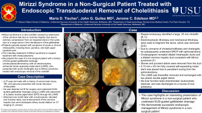
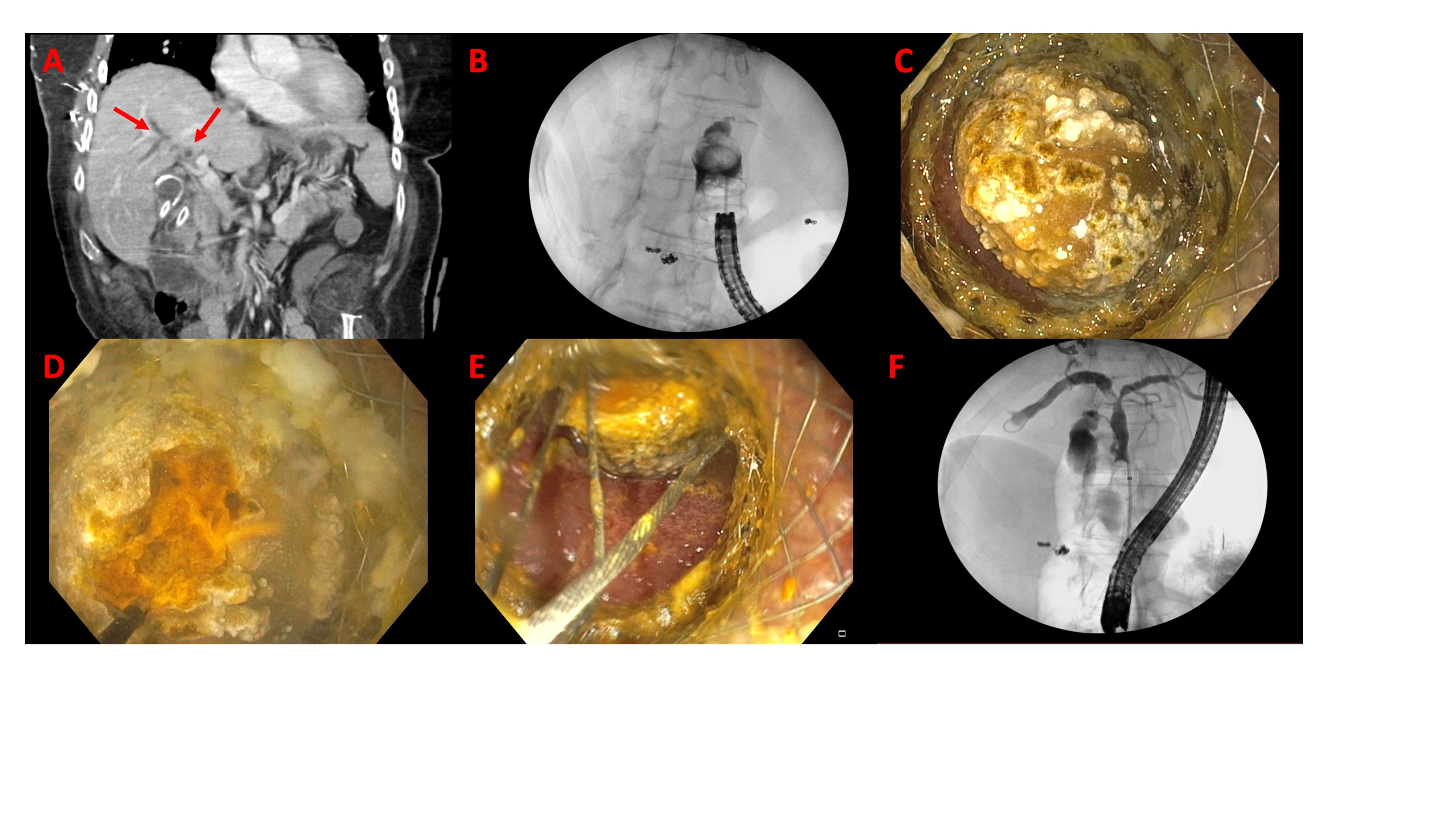
Case Description/Methods: A 37-year-old male with a history of anoxic brain injury, TBI, and seizures presented with acute calculous cholecystitis. He was deemed not fit for surgery and underwent EUS guided drainage using a LAMS with placement of a plastic double pigtail stent (DPS) through the LAMS. He returned one month later with elevated liver function tests, leukocytosis, and enhancement of the common hepatic duct and intrahepatic biliary ductal dilation on CT (A, arrows). He underwent repeat endoscopy, which identified a large 20 mm cholelith (B, C). Electrohydraulic lithotripsy performed under direct visualization and mechanical lithotripsy were used to fragment the stone, which was removed (D,E). He then underwent ERCP with sphincterotomy. Cholangiogram revealed dilated intrahepatic ducts and a dilated common hepatic duct consistent with Mirizzi syndrome (F). Stones and purulent debris were removed from the duct. A 10 mm x 40 mm fully covered self-expanding metal stent was placed due to persistent oozing from the sphincterotomy. The LAMS was then removed and exchanged with two DPS. The patient’s liver function tests and leukocytosis improved and he was discharged on a course of oral antibiotics.
Discussion: This case demonstrates an interesting presentation of Mirizzi syndrome in a non-surgical patient who underwent EUS-guided gallbladder drainage. Successful removal of the cholelith was accomplished using traditional techniques used for removal of choledocholithiasis.

Disclosures:
Maria D. Tischer, BA1, John G. Quiles, MD2, Jerome C. Edelson, MD3. P1080 - Mirizzi Syndrome in a Non-Surgical Patient Treated With Endoscopic Transduodenal Removal of Cholelithiasis, ACG 2024 Annual Scientific Meeting Abstracts. Philadelphia, PA: American College of Gastroenterology.
1Uniformed Services University of the Health Sciences, Bethesda, MD; 2Brooke Army Medical Center, San Antonio, TX; 3Brooke Army Medical Center, Fort Sam Houston, TX
Introduction: Mirizzi syndrome is a rare condition caused by obstruction of the common hepatic duct or common bile duct due to extrinsic compression from an impacted stone in the cystic duct or a large stone in the infundibulum of the gallbladder. Patients typically present with fever, jaundice, and right upper quadrant pain. The mainstay of management is surgery. Here, we present the case of a non-surgical patient with a history of EUS guided gallbladder drainage (cholecystoduodenostomy) with an electrocautery enhanced lumen apposing metal stent (LAMS) who presented with acute cholangitis due to Mirizzi syndrome, treated with endoscopic removal of the cholelith.
Case Description/Methods: A 37-year-old male with a history of anoxic brain injury, TBI, and seizures presented with acute calculous cholecystitis. He was deemed not fit for surgery and underwent EUS guided drainage using a LAMS with placement of a plastic double pigtail stent (DPS) through the LAMS. He returned one month later with elevated liver function tests, leukocytosis, and enhancement of the common hepatic duct and intrahepatic biliary ductal dilation on CT (A, arrows). He underwent repeat endoscopy, which identified a large 20 mm cholelith (B, C). Electrohydraulic lithotripsy performed under direct visualization and mechanical lithotripsy were used to fragment the stone, which was removed (D,E). He then underwent ERCP with sphincterotomy. Cholangiogram revealed dilated intrahepatic ducts and a dilated common hepatic duct consistent with Mirizzi syndrome (F). Stones and purulent debris were removed from the duct. A 10 mm x 40 mm fully covered self-expanding metal stent was placed due to persistent oozing from the sphincterotomy. The LAMS was then removed and exchanged with two DPS. The patient’s liver function tests and leukocytosis improved and he was discharged on a course of oral antibiotics.
Discussion: This case demonstrates an interesting presentation of Mirizzi syndrome in a non-surgical patient who underwent EUS-guided gallbladder drainage. Successful removal of the cholelith was accomplished using traditional techniques used for removal of choledocholithiasis.

Figure: A, arrows: enhancement of the common hepatic duct and intrahepatic biliary ductal dilation on CT; B, C: Cholelith; D,E: Lithotripsy; F: Cholangiogram showing findings consistent with Mirizzi syndrome
Disclosures:
Maria Tischer indicated no relevant financial relationships.
John Quiles indicated no relevant financial relationships.
Jerome Edelson indicated no relevant financial relationships.
Maria D. Tischer, BA1, John G. Quiles, MD2, Jerome C. Edelson, MD3. P1080 - Mirizzi Syndrome in a Non-Surgical Patient Treated With Endoscopic Transduodenal Removal of Cholelithiasis, ACG 2024 Annual Scientific Meeting Abstracts. Philadelphia, PA: American College of Gastroenterology.
