Sunday Poster Session
Category: GI Bleeding
P0763 - Life-Threatening Gastrointestinal Bleeding due to Gastric Dieulafoy Lesion in a Pregnant Woman: A Case Report of a Very Rare Event
Sunday, October 27, 2024
3:30 PM - 7:00 PM ET
Location: Exhibit Hall E

Has Audio

John Thesing, DO
University of Kansas School of Medicine
Wichita, KS
Presenting Author(s)
John Thesing, DO, Stephanie J. Melquist, MD, MPH, Daly Al-Hadeethi, MD, Taylor Jones, DO, William J. Salyers, MD, MPH
University of Kansas School of Medicine, Wichita, KS
Introduction: Upper gastrointestinal bleeding (UGIB) in pregnant women is rarely reported and not well studied. Dieulafoy lesions (DL) are a rare cause of UGIB overall and rarely seen before age 50. We report a rare case of UGIB in a pregnant woman caused by a DL.
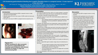
Case Description/Methods: A 27-year-old pregnant woman, G2P1 at 31 weeks gestation, developed sudden onset nausea with massive hematemesis and orthostasis so called EMS. She had 3 subsequent large bloody emesis enroute. On arrival to ER, Hb was 10.3 g/dL with normal blood pressure, platelet count, INR and creatinine. She underwent expectant management with antiemetics, fluids, high dose PPI and was admitted for stabilization and fetal monitoring. Overnight, she experienced more hematemesis, maternal arrhythmia, and evidence of fetal distress so was taken to the OR for emergent EGD. Hb was 7.6 g/dL. A large amount of clotted blood was found in the fundus and body of the stomach. Mucosal tears, possibly due to vomiting, were noted in the fundus and the largest was treated with epinephrine and clipped closed. Due to hemodynamic instability, interventional radiology performed empiric embolization of the left gastroepiploic and left and short gastric arteries as the stomach could not be fully cleared. She rebled on hospital day (HD) 3, Hb now 6.4 g/dL, CT angiography (CTA) was performed showing a tortuous collection of vessels consistent with angiodysplasia in the posterior fundus. On HD 4, she was taken for urgent c-section at 32 weeks and delivered a healthy baby. On HD 7, repeat EGD again revealed a large amount of clotted blood in the fundus. This was cleared revealing a large vessel protruding through the gastric wall consistent with a DL in the area of the angiodysplasia seen on CT (Figure 1). This was treated with epinephrine, and clipped with successful hemostasis. Mom was discharged on HD 9 and baby on HD 30 in good health
Discussion: Life threatening UGIB in pregnant women is rarely reported. Increased portal hypertension due to the gravid uterus is one risk for UGIB, particularly with the vascular malformation seen on CTA in our case. This brings to light the multidisciplinary efforts needed to care for pregnant women with UGIB in order to save the life of mother and baby, as well as the need to carefully examine for DL in those with increased portal hypertension. While CTA is generally avoided during pregnancy, it should be considered as a lifesaving tool in certain circumstances.

Disclosures:
John Thesing, DO, Stephanie J. Melquist, MD, MPH, Daly Al-Hadeethi, MD, Taylor Jones, DO, William J. Salyers, MD, MPH. P0763 - Life-Threatening Gastrointestinal Bleeding due to Gastric Dieulafoy Lesion in a Pregnant Woman: A Case Report of a Very Rare Event, ACG 2024 Annual Scientific Meeting Abstracts. Philadelphia, PA: American College of Gastroenterology.
University of Kansas School of Medicine, Wichita, KS
Introduction: Upper gastrointestinal bleeding (UGIB) in pregnant women is rarely reported and not well studied. Dieulafoy lesions (DL) are a rare cause of UGIB overall and rarely seen before age 50. We report a rare case of UGIB in a pregnant woman caused by a DL.
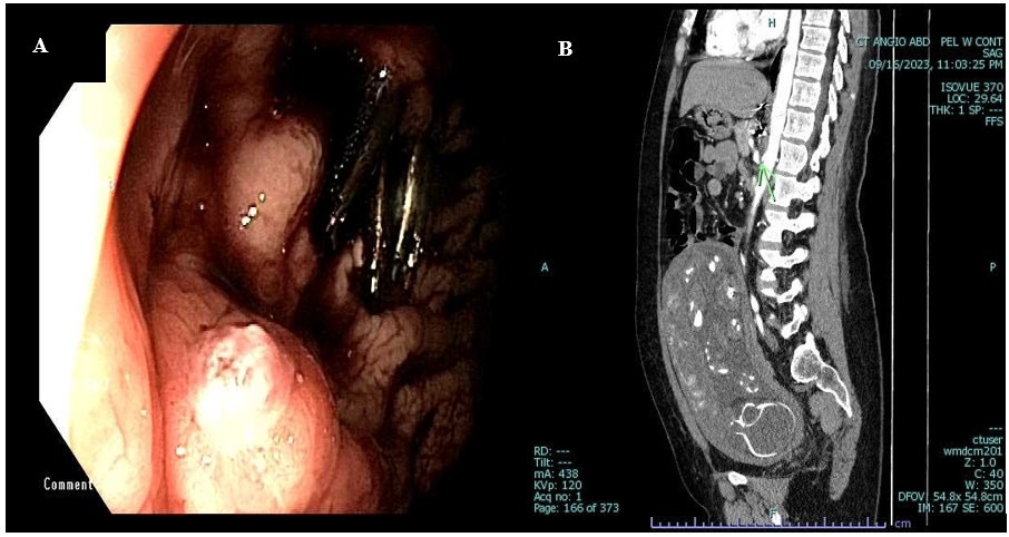
Case Description/Methods: A 27-year-old pregnant woman, G2P1 at 31 weeks gestation, developed sudden onset nausea with massive hematemesis and orthostasis so called EMS. She had 3 subsequent large bloody emesis enroute. On arrival to ER, Hb was 10.3 g/dL with normal blood pressure, platelet count, INR and creatinine. She underwent expectant management with antiemetics, fluids, high dose PPI and was admitted for stabilization and fetal monitoring. Overnight, she experienced more hematemesis, maternal arrhythmia, and evidence of fetal distress so was taken to the OR for emergent EGD. Hb was 7.6 g/dL. A large amount of clotted blood was found in the fundus and body of the stomach. Mucosal tears, possibly due to vomiting, were noted in the fundus and the largest was treated with epinephrine and clipped closed. Due to hemodynamic instability, interventional radiology performed empiric embolization of the left gastroepiploic and left and short gastric arteries as the stomach could not be fully cleared. She rebled on hospital day (HD) 3, Hb now 6.4 g/dL, CT angiography (CTA) was performed showing a tortuous collection of vessels consistent with angiodysplasia in the posterior fundus. On HD 4, she was taken for urgent c-section at 32 weeks and delivered a healthy baby. On HD 7, repeat EGD again revealed a large amount of clotted blood in the fundus. This was cleared revealing a large vessel protruding through the gastric wall consistent with a DL in the area of the angiodysplasia seen on CT (Figure 1). This was treated with epinephrine, and clipped with successful hemostasis. Mom was discharged on HD 9 and baby on HD 30 in good health
Discussion: Life threatening UGIB in pregnant women is rarely reported. Increased portal hypertension due to the gravid uterus is one risk for UGIB, particularly with the vascular malformation seen on CTA in our case. This brings to light the multidisciplinary efforts needed to care for pregnant women with UGIB in order to save the life of mother and baby, as well as the need to carefully examine for DL in those with increased portal hypertension. While CTA is generally avoided during pregnancy, it should be considered as a lifesaving tool in certain circumstances.

Figure: Figure 1. A-Dieulafoy lesion seen in gastric fundus during repeat EGD for UGIB. B- Sagital view of CTA abdomen/pelvis showing vascular malformation in the posterior gastric fundus, marked by arrow.
Disclosures:
John Thesing indicated no relevant financial relationships.
Stephanie Melquist indicated no relevant financial relationships.
Daly Al-Hadeethi indicated no relevant financial relationships.
Taylor Jones indicated no relevant financial relationships.
William Salyers indicated no relevant financial relationships.
John Thesing, DO, Stephanie J. Melquist, MD, MPH, Daly Al-Hadeethi, MD, Taylor Jones, DO, William J. Salyers, MD, MPH. P0763 - Life-Threatening Gastrointestinal Bleeding due to Gastric Dieulafoy Lesion in a Pregnant Woman: A Case Report of a Very Rare Event, ACG 2024 Annual Scientific Meeting Abstracts. Philadelphia, PA: American College of Gastroenterology.
