Sunday Poster Session
Category: Esophagus
P0605 - Masquerading Esophageal Lesions With Diametrically Different Management
Sunday, October 27, 2024
3:30 PM - 7:00 PM ET
Location: Exhibit Hall E

Has Audio

Brianna Hamilton, MD, MS
Houston Methodist Hospital
Houston, TX
Presenting Author(s)
Brianna Hamilton, MD, MS1, Ramon O. Minjares, MD1, Mary R. Schwartz, MD1, Rachel Schiesser, MD2
1Houston Methodist Hospital, Houston, TX; 2Underwood Center for Digestive Disorders, Houston Methodist Hospital, Houston, TX
Introduction: White esophageal lesions are commonly seen and not always biopsied. While some lesions do not require biopsy, others are dependent on pathologic evaluation for diagnosis and appropriate treatment. Both premalignant and benign conditions may be found and can be difficult to distinguish both endoscopically and pathologically.
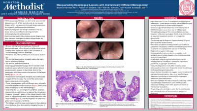
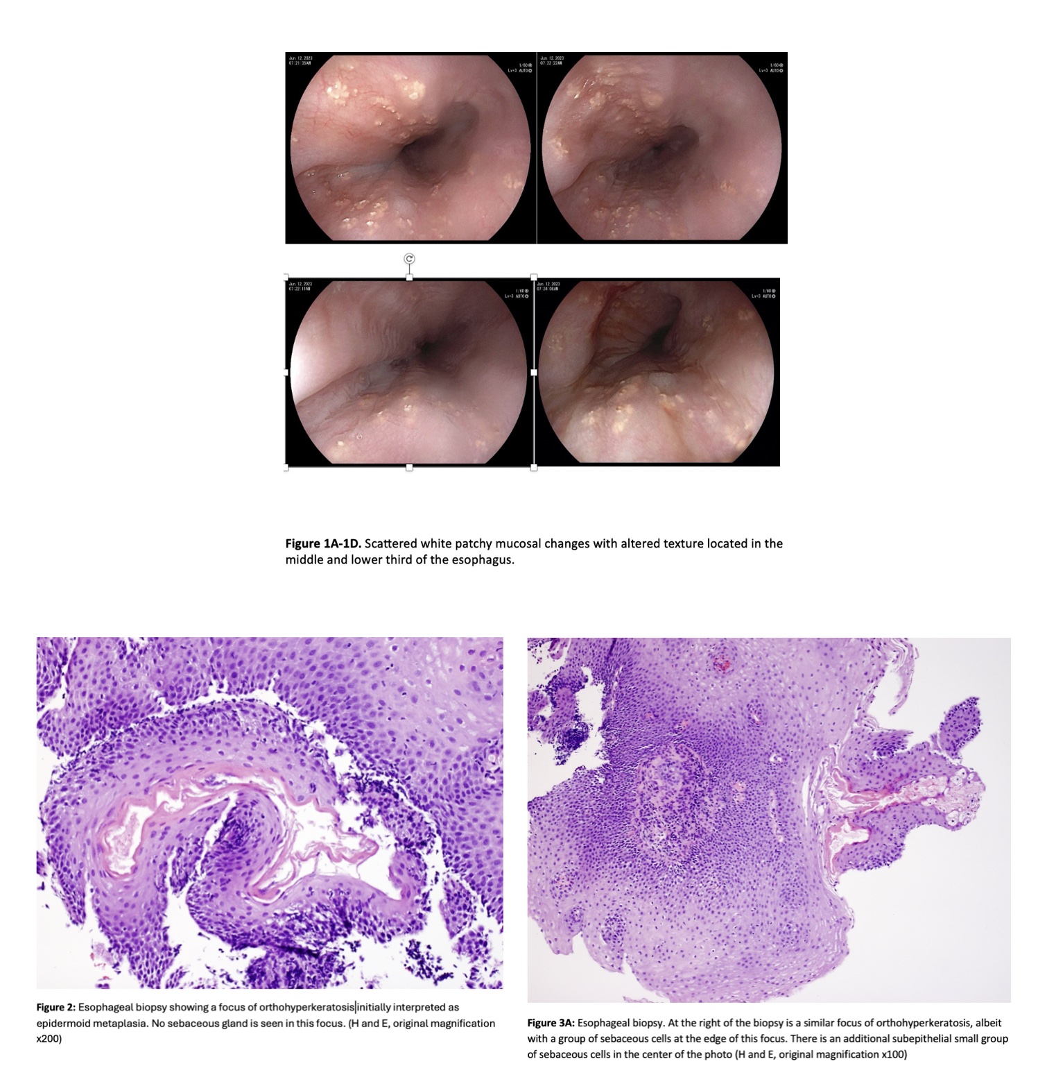
Case Description/Methods: A 60 year old man with a history of gastroesophageal reflux disease on daily pantoprazole presented with “throat-clearing” without dysphagia.The patient underwent elective esophagogastroduodenoscopy which revealed small scattered patchy whitish lesions predominantly located in the middle third of the esophagus with additional lesions in the lower third. These lesions were slightly elevated and unable to be scraped off the mucosa. Tissue biopsies taken with cold forceps were originally reported as epidermoid metaplasia. Given that this is a premalignant condition, ablation, and periodic surveillance were planned for the patient. However, on expert review of the tissue biopsies focal areas of orthokeratosis with associated sebaceous cells were noted, and the diagnosis of sebaceous gland heterotopia was rendered. This benign condition does not require further ablation or surveillance, thus dramatically changing the plan of care.
Discussion: Here we present a case of esophageal sebaceous gland heterotopia, a rare and benign condition. It has been postulated that it arises from either an acquired metaplastic process or a congenital abnormality. The average age at diagnosis is 50 years with no gender predominance. Two-thirds of patients present due to reflux or symptoms of dyspepsia; whereas the remaining one-third of patients are asymptomatic and are incidentally diagnosed via upper endoscopy. Endoscopically it presents as non-scrapable small yellowish-white plaques in the middle and lower third of the esophagus. Esophageal sebaceous gland heterotopia may be misdiagnosed as Candidiasis, xanthomas, squamous papillomas, or glycogen acanthoses. Given the lack of evidence to suggest an increased risk of malignant transformation, there is no benefit of post-diagnostic monitoring or treatment for esophageal sebaceous gland heterotopia. This case highlights the importance of conducting careful histopathologic examinations and correlation with endoscopic findings when white lesions are found on macroscopic evaluation. Proper diagnosis can help to avoid the financial burden and emotional stress of unnecessary or aggressive treatment.

Disclosures:
Brianna Hamilton, MD, MS1, Ramon O. Minjares, MD1, Mary R. Schwartz, MD1, Rachel Schiesser, MD2. P0605 - Masquerading Esophageal Lesions With Diametrically Different Management, ACG 2024 Annual Scientific Meeting Abstracts. Philadelphia, PA: American College of Gastroenterology.
1Houston Methodist Hospital, Houston, TX; 2Underwood Center for Digestive Disorders, Houston Methodist Hospital, Houston, TX
Introduction: White esophageal lesions are commonly seen and not always biopsied. While some lesions do not require biopsy, others are dependent on pathologic evaluation for diagnosis and appropriate treatment. Both premalignant and benign conditions may be found and can be difficult to distinguish both endoscopically and pathologically.
Case Description/Methods: A 60 year old man with a history of gastroesophageal reflux disease on daily pantoprazole presented with “throat-clearing” without dysphagia.The patient underwent elective esophagogastroduodenoscopy which revealed small scattered patchy whitish lesions predominantly located in the middle third of the esophagus with additional lesions in the lower third. These lesions were slightly elevated and unable to be scraped off the mucosa. Tissue biopsies taken with cold forceps were originally reported as epidermoid metaplasia. Given that this is a premalignant condition, ablation, and periodic surveillance were planned for the patient. However, on expert review of the tissue biopsies focal areas of orthokeratosis with associated sebaceous cells were noted, and the diagnosis of sebaceous gland heterotopia was rendered. This benign condition does not require further ablation or surveillance, thus dramatically changing the plan of care.
Discussion: Here we present a case of esophageal sebaceous gland heterotopia, a rare and benign condition. It has been postulated that it arises from either an acquired metaplastic process or a congenital abnormality. The average age at diagnosis is 50 years with no gender predominance. Two-thirds of patients present due to reflux or symptoms of dyspepsia; whereas the remaining one-third of patients are asymptomatic and are incidentally diagnosed via upper endoscopy. Endoscopically it presents as non-scrapable small yellowish-white plaques in the middle and lower third of the esophagus. Esophageal sebaceous gland heterotopia may be misdiagnosed as Candidiasis, xanthomas, squamous papillomas, or glycogen acanthoses. Given the lack of evidence to suggest an increased risk of malignant transformation, there is no benefit of post-diagnostic monitoring or treatment for esophageal sebaceous gland heterotopia. This case highlights the importance of conducting careful histopathologic examinations and correlation with endoscopic findings when white lesions are found on macroscopic evaluation. Proper diagnosis can help to avoid the financial burden and emotional stress of unnecessary or aggressive treatment.

Figure: Figure 1A-1D: Scattered white patchy mucosal changes with altered texture located in the middle and lower third of the esophagus
Figure 2:Esophageal biopsy showing a focus of orthohyperkeratosis initially interpreted as epidermoid metaplasia. No sebaceous gland is seen in this focus. (H and E, original magnification x200)
Figure 3A: Esophageal biopsy. At the right of the biopsy is a similar focus of orthohyperkeratosis, albeit with a group of sebaceous cells at the edge of this focus. There is an additional subepithelial small group of sebaceous cells in the center of the photo (H and E, original magnification x100)
Disclosures:
Brianna Hamilton indicated no relevant financial relationships.
Ramon O. Minjares indicated no relevant financial relationships.
Mary Schwartz indicated no relevant financial relationships.
Rachel Schiesser indicated no relevant financial relationships.
Brianna Hamilton, MD, MS1, Ramon O. Minjares, MD1, Mary R. Schwartz, MD1, Rachel Schiesser, MD2. P0605 - Masquerading Esophageal Lesions With Diametrically Different Management, ACG 2024 Annual Scientific Meeting Abstracts. Philadelphia, PA: American College of Gastroenterology.
