Sunday Poster Session
Category: Esophagus
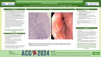
P0606 - A Rare Case of an Atypical Heterotopic Gastric Mucosa of the Lower Esophagus in a Young Man With a Symptomatic Helicobacter pylori Infection
Sunday, October 27, 2024
3:30 PM - 7:00 PM ET
Location: Exhibit Hall E

Has Audio

Mohamed K. Osman, MD, MPH
NYC Health + Hospitals/Harlem
New York, NY
Presenting Author(s)
Mohamed K.. Osman, MD, MPH, Marina Landa, MD, Alvaro Genao, MD, Joan Culpepper-Morgan, MD, FACG
NYC Health + Hospitals/Harlem, New York, NY
Introduction: Heterotopic gastric mucosa (HGM) of the esophagus, also known as inlet patch, are salmon-colored islands of columnar epithelium and are usually found in the cervical esophagus. Goblet cells are not seen. This is in contrast to Barrett’s esophagus (BE), which is characterized by salmon-colored islands or tongues of specialized columnar epithelium in the lower esophagus. This intestinal metaplasia may become dysplastic and lead to cancer. Some studies have linked HGM to BE, while others contend that HGM is likely congenital and BE is acquired because of gastroesophageal reflux (GERD). HGM may be asymptomatic or may be associated with symptoms of laryngopharyngeal reflux. Helicobacter pylori infection (HPI) of HGM is not uncommon. We describe a young man presenting with HGM of the distal esophagus associated HPI infection.
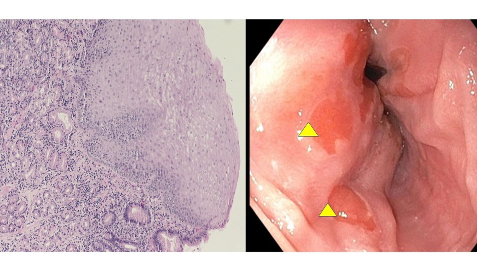
Case Description/Methods: A 21-year-old Hispanic man was diagnosed with chronic adenotonsillitis, allergic rhinitis, and laryngopharyngeal reflux 6 months prior to presentation. He noted heartburn, sore throat, and chest discomfort for 1 year that worsened over the 7 months before presentation. He denied alcohol and tobacco use. Physical was unremarkable. Esophagogastroduodenoscopy showed LA grade B esophagitis and a small hiatal hernia. There were 2 islands of pink mucosa in the lower third of the esophagus (Figure 1). Random gastric biopsies revealed chronic active H. pylori gastritis. No intestinal metaplasia or dysplasia was seen. Lower esophageal biopsy showed columnar cardiac-type mucosa with chronic active H. pylori gastritis, and adjacent squamous mucosa within normal limits. Mid-esophagus biopsy showed normal squamous mucosa. His symptoms resolved after 2 weeks of amoxicillin, clarithromycin, and pantoprazole. Negative HPI stool Ag confirmed eradication 1 month later.
Discussion: HGM occurs mostly in the cervical esophagus. Our case presented with distal esophageal HGM. In addition, he presented with laryngopharyngeal reflux that responded to conventional HPI triple therapy. His history of atopy correlates with a diagnosis of HGM and not Barrett’s. HGM of the upper esophagus is thought to be due to the persistence of embryogenic gastric columnar epithelium. HGM in the distal esophagus has been described in older patients with GERD, median age 59 years. Although HGM at the distal esophagus of this patient might be a complication of GERD and a precursor for Barrett’s, his young age makes a congenital distal HGM patch a more favorable hypothesis.

Disclosures:
Mohamed K.. Osman, MD, MPH, Marina Landa, MD, Alvaro Genao, MD, Joan Culpepper-Morgan, MD, FACG. P0606 - A Rare Case of an Atypical Heterotopic Gastric Mucosa of the Lower Esophagus in a Young Man With a Symptomatic <i>Helicobacter pylori</i> Infection, ACG 2024 Annual Scientific Meeting Abstracts. Philadelphia, PA: American College of Gastroenterology.
NYC Health + Hospitals/Harlem, New York, NY
Introduction: Heterotopic gastric mucosa (HGM) of the esophagus, also known as inlet patch, are salmon-colored islands of columnar epithelium and are usually found in the cervical esophagus. Goblet cells are not seen. This is in contrast to Barrett’s esophagus (BE), which is characterized by salmon-colored islands or tongues of specialized columnar epithelium in the lower esophagus. This intestinal metaplasia may become dysplastic and lead to cancer. Some studies have linked HGM to BE, while others contend that HGM is likely congenital and BE is acquired because of gastroesophageal reflux (GERD). HGM may be asymptomatic or may be associated with symptoms of laryngopharyngeal reflux. Helicobacter pylori infection (HPI) of HGM is not uncommon. We describe a young man presenting with HGM of the distal esophagus associated HPI infection.
Case Description/Methods: A 21-year-old Hispanic man was diagnosed with chronic adenotonsillitis, allergic rhinitis, and laryngopharyngeal reflux 6 months prior to presentation. He noted heartburn, sore throat, and chest discomfort for 1 year that worsened over the 7 months before presentation. He denied alcohol and tobacco use. Physical was unremarkable. Esophagogastroduodenoscopy showed LA grade B esophagitis and a small hiatal hernia. There were 2 islands of pink mucosa in the lower third of the esophagus (Figure 1). Random gastric biopsies revealed chronic active H. pylori gastritis. No intestinal metaplasia or dysplasia was seen. Lower esophageal biopsy showed columnar cardiac-type mucosa with chronic active H. pylori gastritis, and adjacent squamous mucosa within normal limits. Mid-esophagus biopsy showed normal squamous mucosa. His symptoms resolved after 2 weeks of amoxicillin, clarithromycin, and pantoprazole. Negative HPI stool Ag confirmed eradication 1 month later.
Discussion: HGM occurs mostly in the cervical esophagus. Our case presented with distal esophageal HGM. In addition, he presented with laryngopharyngeal reflux that responded to conventional HPI triple therapy. His history of atopy correlates with a diagnosis of HGM and not Barrett’s. HGM of the upper esophagus is thought to be due to the persistence of embryogenic gastric columnar epithelium. HGM in the distal esophagus has been described in older patients with GERD, median age 59 years. Although HGM at the distal esophagus of this patient might be a complication of GERD and a precursor for Barrett’s, his young age makes a congenital distal HGM patch a more favorable hypothesis.

Figure: Islands of salmon-colored mucosa (arrows) at the lower part of the esophagus above the Z-line. Histology with cardiac mucosa with chronic active gastritis, and no intestinal metaplasia (no goblet cells) to the left and normal squamous mucosa to the right.
Disclosures:
Mohamed Osman indicated no relevant financial relationships.
Marina Landa indicated no relevant financial relationships.
Alvaro Genao indicated no relevant financial relationships.
Joan Culpepper-Morgan indicated no relevant financial relationships.
Mohamed K.. Osman, MD, MPH, Marina Landa, MD, Alvaro Genao, MD, Joan Culpepper-Morgan, MD, FACG. P0606 - A Rare Case of an Atypical Heterotopic Gastric Mucosa of the Lower Esophagus in a Young Man With a Symptomatic <i>Helicobacter pylori</i> Infection, ACG 2024 Annual Scientific Meeting Abstracts. Philadelphia, PA: American College of Gastroenterology.
