Sunday Poster Session
Category: Esophagus
P0559 - An Interesting Case of Lymphocytic Esophagitis In Association With Pityriasis Lichenoides Chronica and Rheumatoid Arthritis
Sunday, October 27, 2024
3:30 PM - 7:00 PM ET
Location: Exhibit Hall E

Has Audio
- RM
Raakhi Menon, DO
University of Texas Medical Branch
Galveston, TX
Presenting Author(s)
Raakhi Menon, DO, Kian Abdul-Baki, DO, Kanana Aburayyan, MD, Harshwardhan Thaker, MD, Yamam Al-Saadi, MD
University of Texas Medical Branch, Galveston, TX
Introduction: Lymphocytic esophagitis (LyE) is a rare esophageal disorder of unknown etiology, characterized by dense peripapillary lymphocytes without neutrophils or eosinophils, and spongiosis. Patients typically present with symptoms such as dysphagia or chest pain. Here, we describe a notable case of lymphocytic esophagitis in a patient who presented with food impaction.
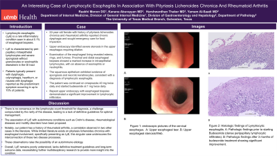
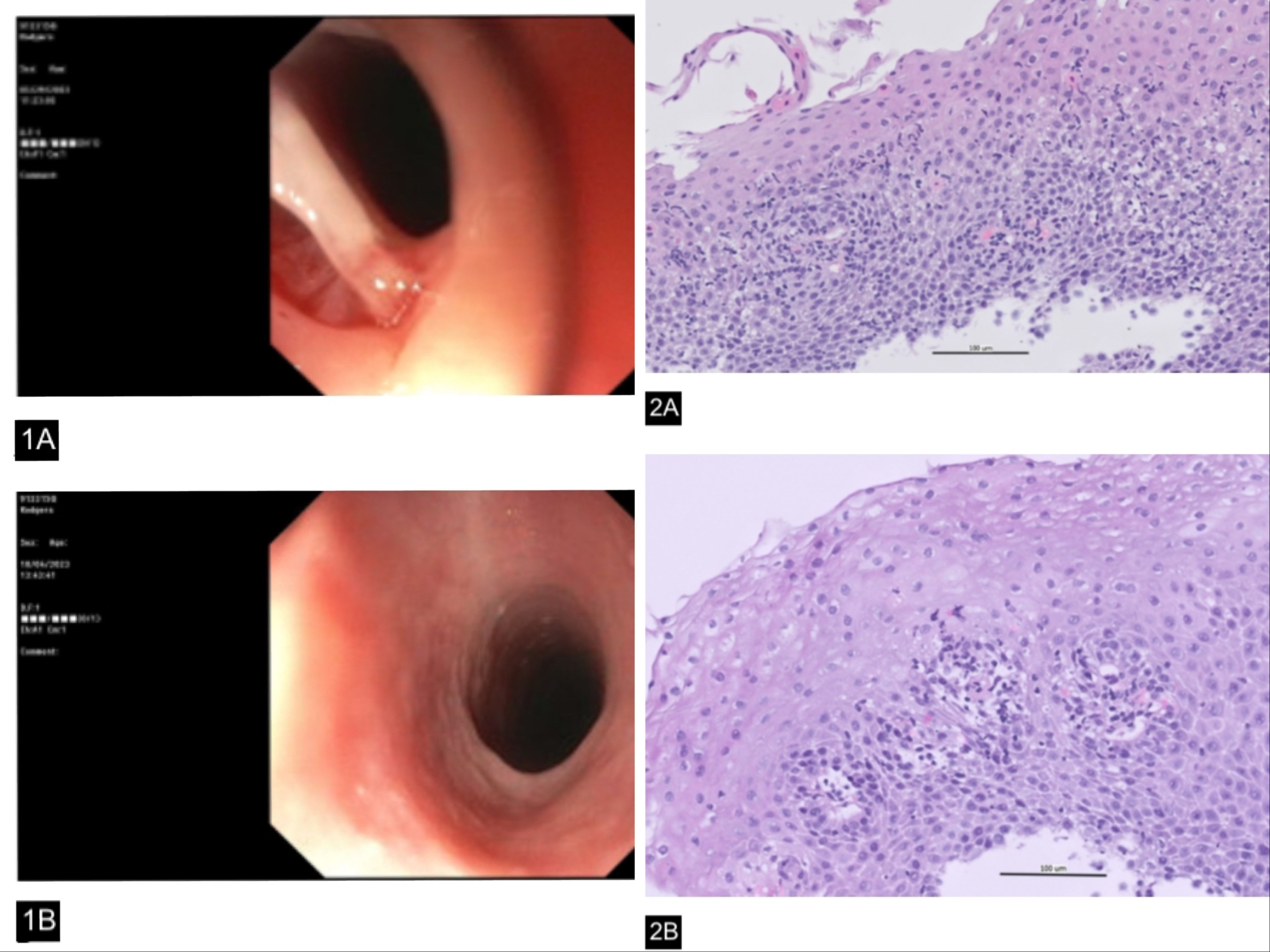
Case Description/Methods: A 53-year-old female with a history of pityriasis lichenoides chronica and rheumatoid arthritis reported chronic dysphagia and sought emergency care for food impaction. A subsequent upper endoscopy revealed a long submucosal tear in the upper esophagus during intubation (see Figure 1A), leading to the procedure's cessation. A follow-up upper endoscopy nine weeks later, employing an ultra-slim scope, identified severe stenosis in the upper esophagus (see Figure 1B). Dilation was performed using a Savary dilator. Examination of the esophageal lining revealed edema, rings, and furrows. Proximal and distal esophageal biopsies were obtained, indicating a marked increase in intraepithelial lymphocytes, with an absence of eosinophils or neutrophils. The squamous epithelium exhibited evidence of spongiosis and necrotic keratinocytes, consistent with a diagnosis of lymphocytic esophagitis. The patient continued omeprazole 40 mg twice daily and started budesonide at 1 mg twice daily. A repeat upper endoscopy with esophageal biopsies demonstrated a significant improvement in lymphocytic infiltration (see Figure 2A-B).
Discussion: LyE typically manifests in females during their 5th-6th decades, presenting a notable contrast to eosinophilic esophagitis (EOE), which predominantly affects males in their 2nd-3rd decades. However, the endoscopic appearance of LyE often mirrors that of EOE. Studies suggest potential associations between LyE and Crohn's disease in children, as well as primary esophageal dysmotility in adults. Notably, our patient has a history of rheumatoid arthritis, a correlation observed in two other cases in the literature. While limited literature exists on pityriasis lichenoides chronica with esophageal involvement, specifically presenting as LyE, this singular case underscores the interconnection of these two disease processes. These observations raise the possibility of an autoimmune etiology. There is no consensus on the lymphocyte count threshold for diagnosis, a challenge compounded by the rarity of the disease, leading to a lack of definitive guidelines for optimal management.

Disclosures:
Raakhi Menon, DO, Kian Abdul-Baki, DO, Kanana Aburayyan, MD, Harshwardhan Thaker, MD, Yamam Al-Saadi, MD. P0559 - An Interesting Case of Lymphocytic Esophagitis In Association With Pityriasis Lichenoides Chronica and Rheumatoid Arthritis, ACG 2024 Annual Scientific Meeting Abstracts. Philadelphia, PA: American College of Gastroenterology.
University of Texas Medical Branch, Galveston, TX
Introduction: Lymphocytic esophagitis (LyE) is a rare esophageal disorder of unknown etiology, characterized by dense peripapillary lymphocytes without neutrophils or eosinophils, and spongiosis. Patients typically present with symptoms such as dysphagia or chest pain. Here, we describe a notable case of lymphocytic esophagitis in a patient who presented with food impaction.
Case Description/Methods: A 53-year-old female with a history of pityriasis lichenoides chronica and rheumatoid arthritis reported chronic dysphagia and sought emergency care for food impaction. A subsequent upper endoscopy revealed a long submucosal tear in the upper esophagus during intubation (see Figure 1A), leading to the procedure's cessation. A follow-up upper endoscopy nine weeks later, employing an ultra-slim scope, identified severe stenosis in the upper esophagus (see Figure 1B). Dilation was performed using a Savary dilator. Examination of the esophageal lining revealed edema, rings, and furrows. Proximal and distal esophageal biopsies were obtained, indicating a marked increase in intraepithelial lymphocytes, with an absence of eosinophils or neutrophils. The squamous epithelium exhibited evidence of spongiosis and necrotic keratinocytes, consistent with a diagnosis of lymphocytic esophagitis. The patient continued omeprazole 40 mg twice daily and started budesonide at 1 mg twice daily. A repeat upper endoscopy with esophageal biopsies demonstrated a significant improvement in lymphocytic infiltration (see Figure 2A-B).
Discussion: LyE typically manifests in females during their 5th-6th decades, presenting a notable contrast to eosinophilic esophagitis (EOE), which predominantly affects males in their 2nd-3rd decades. However, the endoscopic appearance of LyE often mirrors that of EOE. Studies suggest potential associations between LyE and Crohn's disease in children, as well as primary esophageal dysmotility in adults. Notably, our patient has a history of rheumatoid arthritis, a correlation observed in two other cases in the literature. While limited literature exists on pityriasis lichenoides chronica with esophageal involvement, specifically presenting as LyE, this singular case underscores the interconnection of these two disease processes. These observations raise the possibility of an autoimmune etiology. There is no consensus on the lymphocyte count threshold for diagnosis, a challenge compounded by the rarity of the disease, leading to a lack of definitive guidelines for optimal management.

Figure: Figure 1: endoscopic pictures of the cervical esophagus.
A: Upper esophageal tear. B: Upper esophageal stenosis/Web.
Figure 2: Histologic findings of Lymphocytic esophagitis.
A: Pathologic findings prior to starting Budesonide (dense peripapillary lymphocytic infiltrates). B: Pathologic findings after 3 months of Budesonide treatment showing significant improvement.
Disclosures:
Raakhi Menon indicated no relevant financial relationships.
Kian Abdul-Baki indicated no relevant financial relationships.
Kanana Aburayyan indicated no relevant financial relationships.
Harshwardhan Thaker indicated no relevant financial relationships.
Yamam Al-Saadi indicated no relevant financial relationships.
Raakhi Menon, DO, Kian Abdul-Baki, DO, Kanana Aburayyan, MD, Harshwardhan Thaker, MD, Yamam Al-Saadi, MD. P0559 - An Interesting Case of Lymphocytic Esophagitis In Association With Pityriasis Lichenoides Chronica and Rheumatoid Arthritis, ACG 2024 Annual Scientific Meeting Abstracts. Philadelphia, PA: American College of Gastroenterology.
