Sunday Poster Session
Category: Esophagus
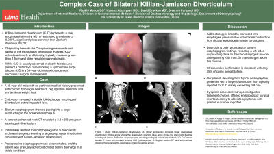
P0561 - Complex Case of Bilateral Killian-Jamieson Diverticulum
Sunday, October 27, 2024
3:30 PM - 7:00 PM ET
Location: Exhibit Hall E

Has Audio
- RM
Raakhi Menon, DO
University of Texas Medical Branch
Galveston, TX
Presenting Author(s)
Raakhi Menon, DO, Ayesha Khan, DO, Kanana Aburayyan, MD, David Bracken, MD, Sreeram Parupudi, MD
University of Texas Medical Branch, Galveston, TX
Introduction: Killian-Jamieson diverticulum (KJD) represents a rare esophageal anomaly, with an estimated prevalence of 0.025%, significantly less common than Zenker's diverticulum (ZD). Originating beneath the Cricopharyngeus muscle and lateral to the esophageal longitudinal muscles, KJD extends anteriorly and laterally, typically measuring less than 1.5 cm and often remaining asymptomatic. While KJD is usually observed in elderly females, we present a distinctive case involving a symptomatic large bilobed KJD in a 39-year-old male who underwent successful surgical management.
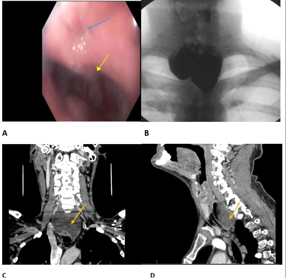
Case Description/Methods: A 39-year-old male with no pertinent medical history presented with chronic dysphagia, heartburn, regurgitation, halitosis, and unintentional weight loss. Endoscopic examination revealed a sizable bilobed upper esophageal diverticulum with a substantial opening, containing copious thick mucus but no impacted food (See Figure 1A). Barium esophagogram demonstrated pooling into a large outpouching in the posterior esophagus, displacing it anteriorly, with intermittent regurgitation into the hypopharynx and esophagus (Figure 1B). A contrast-enhanced neck CT revealed a 3.8 x 6.5 cm upper esophageal diverticulum (Figure 1C-D). The patient was referred to otolaryngology and subsequently underwent surgery, revealing a large bilobed esophageal diverticulum consistent with Killian-Jamieson Diverticulum, measuring 4 cm in neck width, 3 cm in the right lobe, and 6 cm in the left lobe. Open transcervical esophageal diverticulectomy was performed with increased complexity due to the width of the neck and the infra-cricopharyngeal muscle location. Postoperative esophagogram was unremarkable, and the patient was gradually advanced on diet before discharge in a stable condition.
Discussion: KJD's etiology is linked to increased intra-esophageal pressure due to functional obstruction from circular esophageal muscle contractions. Diagnosis is often prompted by barium esophagogram findings, revealing a left-sided outpouching distal to the cricopharyngeal muscle, distinguishing KJD from ZD that emerges above this muscle. Intraoperative confirmation is standard, with only 25% of cases being bilateral. Our patient, deviating from typical demographics, presented with a larger diverticulum than typically reported for KJD (rarely exceeding 3.8 cm). Symptom-dependent management guides treatment choices, offering endoscopic or surgical diverticulectomy to alleviate symptoms, with positive outcomes reported.

Disclosures:
Raakhi Menon, DO, Ayesha Khan, DO, Kanana Aburayyan, MD, David Bracken, MD, Sreeram Parupudi, MD. P0561 - Complex Case of Bilateral Killian-Jamieson Diverticulum, ACG 2024 Annual Scientific Meeting Abstracts. Philadelphia, PA: American College of Gastroenterology.
University of Texas Medical Branch, Galveston, TX
Introduction: Killian-Jamieson diverticulum (KJD) represents a rare esophageal anomaly, with an estimated prevalence of 0.025%, significantly less common than Zenker's diverticulum (ZD). Originating beneath the Cricopharyngeus muscle and lateral to the esophageal longitudinal muscles, KJD extends anteriorly and laterally, typically measuring less than 1.5 cm and often remaining asymptomatic. While KJD is usually observed in elderly females, we present a distinctive case involving a symptomatic large bilobed KJD in a 39-year-old male who underwent successful surgical management.
Case Description/Methods: A 39-year-old male with no pertinent medical history presented with chronic dysphagia, heartburn, regurgitation, halitosis, and unintentional weight loss. Endoscopic examination revealed a sizable bilobed upper esophageal diverticulum with a substantial opening, containing copious thick mucus but no impacted food (See Figure 1A). Barium esophagogram demonstrated pooling into a large outpouching in the posterior esophagus, displacing it anteriorly, with intermittent regurgitation into the hypopharynx and esophagus (Figure 1B). A contrast-enhanced neck CT revealed a 3.8 x 6.5 cm upper esophageal diverticulum (Figure 1C-D). The patient was referred to otolaryngology and subsequently underwent surgery, revealing a large bilobed esophageal diverticulum consistent with Killian-Jamieson Diverticulum, measuring 4 cm in neck width, 3 cm in the right lobe, and 6 cm in the left lobe. Open transcervical esophageal diverticulectomy was performed with increased complexity due to the width of the neck and the infra-cricopharyngeal muscle location. Postoperative esophagogram was unremarkable, and the patient was gradually advanced on diet before discharge in a stable condition.
Discussion: KJD's etiology is linked to increased intra-esophageal pressure due to functional obstruction from circular esophageal muscle contractions. Diagnosis is often prompted by barium esophagogram findings, revealing a left-sided outpouching distal to the cricopharyngeal muscle, distinguishing KJD from ZD that emerges above this muscle. Intraoperative confirmation is standard, with only 25% of cases being bilateral. Our patient, deviating from typical demographics, presented with a larger diverticulum than typically reported for KJD (rarely exceeding 3.8 cm). Symptom-dependent management guides treatment choices, offering endoscopic or surgical diverticulectomy to alleviate symptoms, with positive outcomes reported.

Figure: Figure 1 (A-D): Killian-Jamieson diverticulum.
A: Upper endoscopy showing upper esophageal diverticulum. Yellow arrow shows the diverticulum opening. Blue arrow shows the opening to the true esophageal lumen.
B: Barium esophagogram showing pooling of barium into bilobed KJD.
C: Coronal section CT neck with contrast showing KJD (yellow arrow).
D: Sagittal section CT neck with contrast showing KJD pushing the esophagus anteriorly (yellow arrow).
Disclosures:
Raakhi Menon indicated no relevant financial relationships.
Ayesha Khan indicated no relevant financial relationships.
Kanana Aburayyan indicated no relevant financial relationships.
David Bracken indicated no relevant financial relationships.
Sreeram Parupudi indicated no relevant financial relationships.
Raakhi Menon, DO, Ayesha Khan, DO, Kanana Aburayyan, MD, David Bracken, MD, Sreeram Parupudi, MD. P0561 - Complex Case of Bilateral Killian-Jamieson Diverticulum, ACG 2024 Annual Scientific Meeting Abstracts. Philadelphia, PA: American College of Gastroenterology.
