Sunday Poster Session
Category: Endoscopy Video Forum
P0460 - Role of Red Dichromatic Imaging With Upper GI Bleed
Sunday, October 27, 2024
3:30 PM - 7:00 PM ET
Location: Exhibit Hall E

Has Audio
- PC
Prahan Chetlur, MD
NYU Langone Health
New York, NY
Presenting Author(s)
Prahan Chetlur, MD, Rushi Talati, MD, MBA, Melissa Latorre Debordeaux, MD
NYU Langone Health, New York, NY
Introduction: Red Dichromatic Imaging, or RDI, utilizes red and amber wavelengths (600 nm and 630 nm) to help enhance the color contrast between bleeding points and surrounding pooled blood. Preliminary studies have shown higher rates of bleeding detection in various endoscopic procedures, including for endoscopic submucosal dissection and esophageal varices.
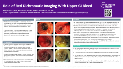
Case Description/Methods: The patient is an elderly woman with a history of metastatic lung carcinoid (on active radiotherapy and chemotherapy) status post left upper lobe lung resection who was initially admitted for MSSA bacteremia. On admission day 8, she was noted to have an episode of maroon stool and hypotension prompting upgrade to the MICU for further evaluation. After initial hemodynamic stabilization, the patient underwent upper endoscopy. On scope insertion, there was a moderate amount of red blood and clot in the antrum that was cleared with copious irrigation. A single 20mm oozing duodenal ulcer (Forrest Class Ib) was found in the duodenal sweep. At this time, RDI was employed to localize the point of bleeding from this ulcer – a focal area that was not well visualized with standard white light imaging (WLI). After triangulation of the culprit lesion, several techniques for hemostasis were deployed. First, 3 mL of epinephrine was injected with improved visualization. Two attempts at over-the-scope clip placement were made but unsuccessful due to extensive granulation tissue at the site of bleeding. Thermal coagulation using a bipolar probe was then performed with successful hemostasis. Lastly, 2 hemostatic sprays were deployed to the cauterized area to prevent future bleeding. After the procedure, both the patient’s hemoglobin and hemodynamics stabilized.
Discussion: In this case, RDI played a key role in managing a challenging duodenal ulcer bleed refractory to epinephrine and over the scope clip placement by facilitating triangulation of the precise area needing thermal coagulation. Here, we were quickly able to identify the site of oozing that was not well-visualized by standard WLI. The success of RDI in this case demonstrates the promise of this technology in evaluating UGIB, particularly in an emergency setting where prompt hemostasis is critical to a favorable patient outcome. Furthermore, it is a useful tool to identify active bleeding sites, particularly for more inexperienced endoscopists. Further studies with standardized RDI versus WLI protocols for upper gastrointestinal bleeding evaluation will help elucidate its routine use in upper endoscopy evaluation.
Disclosures:
Prahan Chetlur, MD, Rushi Talati, MD, MBA, Melissa Latorre Debordeaux, MD. P0460 - Role of Red Dichromatic Imaging With Upper GI Bleed, ACG 2024 Annual Scientific Meeting Abstracts. Philadelphia, PA: American College of Gastroenterology.
NYU Langone Health, New York, NY
Introduction: Red Dichromatic Imaging, or RDI, utilizes red and amber wavelengths (600 nm and 630 nm) to help enhance the color contrast between bleeding points and surrounding pooled blood. Preliminary studies have shown higher rates of bleeding detection in various endoscopic procedures, including for endoscopic submucosal dissection and esophageal varices.
Case Description/Methods: The patient is an elderly woman with a history of metastatic lung carcinoid (on active radiotherapy and chemotherapy) status post left upper lobe lung resection who was initially admitted for MSSA bacteremia. On admission day 8, she was noted to have an episode of maroon stool and hypotension prompting upgrade to the MICU for further evaluation. After initial hemodynamic stabilization, the patient underwent upper endoscopy. On scope insertion, there was a moderate amount of red blood and clot in the antrum that was cleared with copious irrigation. A single 20mm oozing duodenal ulcer (Forrest Class Ib) was found in the duodenal sweep. At this time, RDI was employed to localize the point of bleeding from this ulcer – a focal area that was not well visualized with standard white light imaging (WLI). After triangulation of the culprit lesion, several techniques for hemostasis were deployed. First, 3 mL of epinephrine was injected with improved visualization. Two attempts at over-the-scope clip placement were made but unsuccessful due to extensive granulation tissue at the site of bleeding. Thermal coagulation using a bipolar probe was then performed with successful hemostasis. Lastly, 2 hemostatic sprays were deployed to the cauterized area to prevent future bleeding. After the procedure, both the patient’s hemoglobin and hemodynamics stabilized.
Discussion: In this case, RDI played a key role in managing a challenging duodenal ulcer bleed refractory to epinephrine and over the scope clip placement by facilitating triangulation of the precise area needing thermal coagulation. Here, we were quickly able to identify the site of oozing that was not well-visualized by standard WLI. The success of RDI in this case demonstrates the promise of this technology in evaluating UGIB, particularly in an emergency setting where prompt hemostasis is critical to a favorable patient outcome. Furthermore, it is a useful tool to identify active bleeding sites, particularly for more inexperienced endoscopists. Further studies with standardized RDI versus WLI protocols for upper gastrointestinal bleeding evaluation will help elucidate its routine use in upper endoscopy evaluation.
Disclosures:
Prahan Chetlur indicated no relevant financial relationships.
Rushi Talati indicated no relevant financial relationships.
Melissa Latorre Debordeaux: Enterasense – Consultant. Medtronic – Consultant. Motus GI – Consultant. Olympus – Consultant. OVESCO – Consultant. VTI – Consultant.
Prahan Chetlur, MD, Rushi Talati, MD, MBA, Melissa Latorre Debordeaux, MD. P0460 - Role of Red Dichromatic Imaging With Upper GI Bleed, ACG 2024 Annual Scientific Meeting Abstracts. Philadelphia, PA: American College of Gastroenterology.
