Sunday Poster Session
Category: Endoscopy Video Forum
P0465 - Successful Closure of a Delayed Persistent Duodenal Perforation Using Endoscopic Suturing After Failed Surgical Intervention
Sunday, October 27, 2024
3:30 PM - 7:00 PM ET
Location: Exhibit Hall E

Has Audio

Jameel Alp, MD
University of Minnesota
Minnetonka, MN
Presenting Author(s)
Jameel Alp, MD1, Rahul Karna, MD2, Natalie Wilson, MD2, Mohammad Bilal, MD3
1University of Minnesota, Minnetonka, MN; 2University of Minnesota, Minneapolis, MN; 3University of Minnesota and Minneapolis VA Health Care System, Minneapolis, MN
Introduction: Delayed duodenal perforations are challenging to manage endoscopically due to ulcer fibrosis and the angulations of the duodenum. However, with advances in endoscopic techniques, endoscopic closure can be possible. Here we present a case of successful endoscopic management of a delayed duodenal perforation using a full-thickness suturing device.
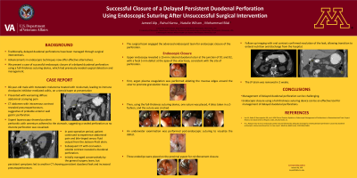
Case Description/Methods: An 80-year-old man with metastatic melanoma on nivolumab and immune checkpoint inhibitor colitis on steroid therapy presented with abdominal pain. Computed tomography scan revealed pneumoperitoneum and suspected perforation on the anterior gastroduodenal wall. Urgent laparoscopy was performed which showed purulent peritonitis with omentum adhering to the gastroduodenal wall, but no obvious perforation was seen. A Jackson-Pratt (JP) drain was placed. Post-operatively, the patient had severe pain and bile-tinged serous fluid from the JP drain. A subsequent computed tomography scan with oral contrast showed a persistent duodenal leak and increased pneumoperitoneum. The patient was referred to advanced endoscopy, and an esophagogastroduodenoscopy was performed which revealed a 15 mm duodenal ulcer at the junction of the 1st and 2nd portion of the duodenum, with a focal defect at the apex of the ulcer consistent with a full-thickness perforation. Decision was made to attempt endoscopic closure. Argon plasma coagulation was used to ablate ulcer edges to promote granulation. Full-thickness endoscopic closure was then performed using the over-the-scope suturing device. Four bites were placed in a “Z” configuration and suture was cinched. Three additional endoclips were placed for reinforcement at the proximal end of the defect. A nasoduodenal tube was placed and connected to low intermittent wall suction. Follow-up imaging with oral contrast did not show any contrast extravasation, and the patient was transitioned to enteral nutrition and discharged. The JP drain was removed after 2 weeks, and patient has not had recurrence of duodenal leak on 6 months follow-up.
Discussion: Our case suggests that endoscopic full-thickness suturing can be effectively used in the duodenum for closure of delayed duodenal perforations and leaks in combination with percutaneous drain placement.
Disclosures:
Jameel Alp, MD1, Rahul Karna, MD2, Natalie Wilson, MD2, Mohammad Bilal, MD3. P0465 - Successful Closure of a Delayed Persistent Duodenal Perforation Using Endoscopic Suturing After Failed Surgical Intervention, ACG 2024 Annual Scientific Meeting Abstracts. Philadelphia, PA: American College of Gastroenterology.
1University of Minnesota, Minnetonka, MN; 2University of Minnesota, Minneapolis, MN; 3University of Minnesota and Minneapolis VA Health Care System, Minneapolis, MN
Introduction: Delayed duodenal perforations are challenging to manage endoscopically due to ulcer fibrosis and the angulations of the duodenum. However, with advances in endoscopic techniques, endoscopic closure can be possible. Here we present a case of successful endoscopic management of a delayed duodenal perforation using a full-thickness suturing device.
Case Description/Methods: An 80-year-old man with metastatic melanoma on nivolumab and immune checkpoint inhibitor colitis on steroid therapy presented with abdominal pain. Computed tomography scan revealed pneumoperitoneum and suspected perforation on the anterior gastroduodenal wall. Urgent laparoscopy was performed which showed purulent peritonitis with omentum adhering to the gastroduodenal wall, but no obvious perforation was seen. A Jackson-Pratt (JP) drain was placed. Post-operatively, the patient had severe pain and bile-tinged serous fluid from the JP drain. A subsequent computed tomography scan with oral contrast showed a persistent duodenal leak and increased pneumoperitoneum. The patient was referred to advanced endoscopy, and an esophagogastroduodenoscopy was performed which revealed a 15 mm duodenal ulcer at the junction of the 1st and 2nd portion of the duodenum, with a focal defect at the apex of the ulcer consistent with a full-thickness perforation. Decision was made to attempt endoscopic closure. Argon plasma coagulation was used to ablate ulcer edges to promote granulation. Full-thickness endoscopic closure was then performed using the over-the-scope suturing device. Four bites were placed in a “Z” configuration and suture was cinched. Three additional endoclips were placed for reinforcement at the proximal end of the defect. A nasoduodenal tube was placed and connected to low intermittent wall suction. Follow-up imaging with oral contrast did not show any contrast extravasation, and the patient was transitioned to enteral nutrition and discharged. The JP drain was removed after 2 weeks, and patient has not had recurrence of duodenal leak on 6 months follow-up.
Discussion: Our case suggests that endoscopic full-thickness suturing can be effectively used in the duodenum for closure of delayed duodenal perforations and leaks in combination with percutaneous drain placement.
Disclosures:
Jameel Alp indicated no relevant financial relationships.
Rahul Karna indicated no relevant financial relationships.
Natalie Wilson indicated no relevant financial relationships.
Mohammad Bilal: Boston Scientific – Consultant. Cook endoscopy – Speakers Bureau.
Jameel Alp, MD1, Rahul Karna, MD2, Natalie Wilson, MD2, Mohammad Bilal, MD3. P0465 - Successful Closure of a Delayed Persistent Duodenal Perforation Using Endoscopic Suturing After Failed Surgical Intervention, ACG 2024 Annual Scientific Meeting Abstracts. Philadelphia, PA: American College of Gastroenterology.
