Sunday Poster Session
Category: Colon
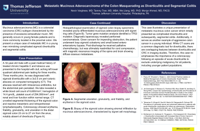
P0386 - Metastatic Mucinous Adenocarcinoma of the Colon Masquerading as Diverticulitis and Segmental Colitis
Sunday, October 27, 2024
3:30 PM - 7:00 PM ET
Location: Exhibit Hall E

Has Audio
- NV
Nevin Varghese, MD
Thomas Jefferson University Hospital
Philadelphia, PA
Presenting Author(s)
Nevin Varghese, MD, Tammy Tran, MD, MBA, Wei Jiang, MD, PhD, Monjur Ahmed, MD, FACG
Thomas Jefferson University Hospital, Philadelphia, PA
Introduction: Mucinous adenocarcinoma (MC) is a colorectal carcinoma CRC subtype characterized by the presence of excessive extracellular mucin. MC generally occurs in young female patients and is more commonly located in the proximal colon. We present a unique case of metastatic MC in a young man mimicking complicated sigmoid diverticulitis and segmental colitis.
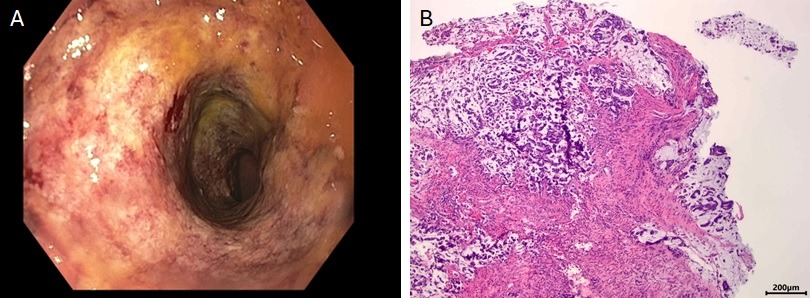
Case Description/Methods: A 32-year-old male with a past medical history of treated chronic hepatitis C and tobacco use presented to the hospital with dull, aching left lower quadrant abdominal pain lasting for three months. Three months prior, he was diagnosed with sigmoid diverticulitis with a 3x3.5 cm peri-colonic abscess on computed tomography (CT). The abscess resolved with intravenous antibiotics, but the abdominal pain persisted. His labs revealed a white blood cell count of 6,600/mm3, hemoglobin of 14.4 gm/dL, platelet count of 294,000/mm3, and renal and liver function within normal range. CT unveiled segmental thickening of the sigmoid colon and reactive mesenteric and retroperitoneal lymphadenopathy. Colonoscopy showed erythema, friability, granularity, and ulceration in the distal sigmoid colon 23 cm to 27 cm from the anus, notably absent of diverticula (Figure A). Histopathological examination of sigmoid colon biopsy specimens revealed poorly differentiated mucinous adenocarcinoma with signet ring cells (Figure B). Tumor gene mutation analysis identified a TP53 mutation. Diagnostic laparoscopy revealed peritoneal carcinomatosis. Given concern for impending obstruction, the patient underwent loop sigmoid colostomy and small bowel entero-enterostomy bypass. Post-discharge he received palliative chemotherapy but was ultimately readmitted for cord compression, with magnetic resonance imaging of the spine and brain showing diffuse osseous metastases.
Discussion: This case illustrates a unique presentation of metastatic mucinous colon cancer which initially presented as complicated diverticulitis and subsequently, segmental colitis. Additionally, it serves as another example of aggressive colon cancer in a young individual. While CT scans are a common diagnostic tool for diverticulitis, there are overlapping features between diverticulitis and CRC in imaging studies. Therefore, this case re-emphasizes the importance of a colonoscopy following an episode of acute diverticulitis to exclude underlying malignancy for all patients, including younger patient populations.

Disclosures:
Nevin Varghese, MD, Tammy Tran, MD, MBA, Wei Jiang, MD, PhD, Monjur Ahmed, MD, FACG. P0386 - Metastatic Mucinous Adenocarcinoma of the Colon Masquerading as Diverticulitis and Segmental Colitis, ACG 2024 Annual Scientific Meeting Abstracts. Philadelphia, PA: American College of Gastroenterology.
Thomas Jefferson University Hospital, Philadelphia, PA
Introduction: Mucinous adenocarcinoma (MC) is a colorectal carcinoma CRC subtype characterized by the presence of excessive extracellular mucin. MC generally occurs in young female patients and is more commonly located in the proximal colon. We present a unique case of metastatic MC in a young man mimicking complicated sigmoid diverticulitis and segmental colitis.
Case Description/Methods: A 32-year-old male with a past medical history of treated chronic hepatitis C and tobacco use presented to the hospital with dull, aching left lower quadrant abdominal pain lasting for three months. Three months prior, he was diagnosed with sigmoid diverticulitis with a 3x3.5 cm peri-colonic abscess on computed tomography (CT). The abscess resolved with intravenous antibiotics, but the abdominal pain persisted. His labs revealed a white blood cell count of 6,600/mm3, hemoglobin of 14.4 gm/dL, platelet count of 294,000/mm3, and renal and liver function within normal range. CT unveiled segmental thickening of the sigmoid colon and reactive mesenteric and retroperitoneal lymphadenopathy. Colonoscopy showed erythema, friability, granularity, and ulceration in the distal sigmoid colon 23 cm to 27 cm from the anus, notably absent of diverticula (Figure A). Histopathological examination of sigmoid colon biopsy specimens revealed poorly differentiated mucinous adenocarcinoma with signet ring cells (Figure B). Tumor gene mutation analysis identified a TP53 mutation. Diagnostic laparoscopy revealed peritoneal carcinomatosis. Given concern for impending obstruction, the patient underwent loop sigmoid colostomy and small bowel entero-enterostomy bypass. Post-discharge he received palliative chemotherapy but was ultimately readmitted for cord compression, with magnetic resonance imaging of the spine and brain showing diffuse osseous metastases.
Discussion: This case illustrates a unique presentation of metastatic mucinous colon cancer which initially presented as complicated diverticulitis and subsequently, segmental colitis. Additionally, it serves as another example of aggressive colon cancer in a young individual. While CT scans are a common diagnostic tool for diverticulitis, there are overlapping features between diverticulitis and CRC in imaging studies. Therefore, this case re-emphasizes the importance of a colonoscopy following an episode of acute diverticulitis to exclude underlying malignancy for all patients, including younger patient populations.

Figure: Figure A. Segmental ulceration, granularity, and friability, and erythema in the sigmoid colon.
Figure B. Biopsy of the sigmoid colon showing stromal infiltration by mucinous adenocarcinoma, characterized by signet cell morphology.
Figure B. Biopsy of the sigmoid colon showing stromal infiltration by mucinous adenocarcinoma, characterized by signet cell morphology.
Disclosures:
Nevin Varghese indicated no relevant financial relationships.
Tammy Tran indicated no relevant financial relationships.
Wei Jiang indicated no relevant financial relationships.
Monjur Ahmed indicated no relevant financial relationships.
Nevin Varghese, MD, Tammy Tran, MD, MBA, Wei Jiang, MD, PhD, Monjur Ahmed, MD, FACG. P0386 - Metastatic Mucinous Adenocarcinoma of the Colon Masquerading as Diverticulitis and Segmental Colitis, ACG 2024 Annual Scientific Meeting Abstracts. Philadelphia, PA: American College of Gastroenterology.
