Sunday Poster Session
Category: Colon
P0388 - Lymphogranuloma Venereum (LGV) in Disguise: A Misleading Case of Proctitis in a Young Patient With HIV
Sunday, October 27, 2024
3:30 PM - 7:00 PM ET
Location: Exhibit Hall E

Has Audio
.jpg)
Maria Teresa Medina Rojas, MD
NYC Health + Hospitals/Jacobi
Brooklyn, NY
Presenting Author(s)
Maria Teresa Medina Rojas, MD1, Carlos Figueredo, MD2, Douglas Simon, MD3, Nichelle Simmons, MD3, Sherrie White, MD3, Donald P. Kotler, MD4
1NYC Health + Hospitals/Jacobi, Brooklyn, NY; 2Montefiore Medical Center, Albert Einstein College of Medicine, Bronx, NY; 3NYC Health + Hospitals/Jacobi, Bronx, NY; 4Albert Einstein College of Medicine, Bronx, NY
Introduction: Gastrointestinal disease in HIV/AIDS does not follow Occam’s razor, the law of parsimony. The evaluation must not stop once a single pathogen is found, as a more significant one might be missed. A clinical-pathologic correlation is vital for diagnosis. LGV has been reported as a cause of proctitis in HIV-positive individuals. We present a case of severe proctitis with multiple potential etiologies.
Case Description/Methods: A 31-year-old HIV-infected bisexual male, poorly adherent to therapy presented with dyspareunia, abdominal pain, intermittent diarrhea, hematochezia, and 20-pound weight loss over 4 months. Physical examination showed a rectal mass. Blood work noted CD4 365/mm3, and VL 467 copies/mL. CT scan showed extensive rectal thickening and prominent presacral, inferior mesenteric, and perirectal adenopathy (figures 1 and 2). A colonoscopy showed a fungating, infiltrative, friable rectal mass, which was biopsied (figure 3). The rest of the colon was uninvolved. Pathology showed acute and chronically inflamed granulation tissue with viral inclusions consistent with CMV and lymphoid hyperplasia but no malignancy (figure 4).
Further evaluation was performed as the presentation was not consistent with CMV colitis. Serum Chlamydia trachomatis IgG was elevated (1:1024) and rectal PCR was positive for Chlamydia. In addition, CMV PCR was 135 IU/mL, and Stool PCR was positive for ETEC/EAEC, but cryptococcus, toxoplasma, and syphilis, were all negative. The diagnosis of LGV was made and doxycycline was started with prompt improvement in symptoms. Repeat sigmoidoscopy 2 weeks later showed persistent CMV inclusions, and valganciclovir was added. Symptoms resolved completely.
Discussion: In our patient, CT scan and colonoscopy findings raised concern for malignancy, while biopsy showed CMV inclusions. However, clinical presentation, CD4 cell count, inflammation limited to the rectum, and lymphocytic hyperplasia on biopsy did not correlate with CMV colitis, but rather LGV. Positive rectal PCR and serum chlamydia titers were pivotal in the diagnosis, as was the response to therapy. The biopsy findings were likely due to CMV reactivation related to severe inflammation. However, because of persistent viral inclusions on repeat biopsy, CMV was treated, despite the major improvement of his symptoms with doxycycline. In conclusion, in an HIV/AIDS patient, it is key to perform a thorough evaluation even after finding a single pathogen to avoid diagnostic delays and achieve better care.

Disclosures:
Maria Teresa Medina Rojas, MD1, Carlos Figueredo, MD2, Douglas Simon, MD3, Nichelle Simmons, MD3, Sherrie White, MD3, Donald P. Kotler, MD4. P0388 - Lymphogranuloma Venereum (LGV) in Disguise: A Misleading Case of Proctitis in a Young Patient With HIV, ACG 2024 Annual Scientific Meeting Abstracts. Philadelphia, PA: American College of Gastroenterology.
1NYC Health + Hospitals/Jacobi, Brooklyn, NY; 2Montefiore Medical Center, Albert Einstein College of Medicine, Bronx, NY; 3NYC Health + Hospitals/Jacobi, Bronx, NY; 4Albert Einstein College of Medicine, Bronx, NY
Introduction: Gastrointestinal disease in HIV/AIDS does not follow Occam’s razor, the law of parsimony. The evaluation must not stop once a single pathogen is found, as a more significant one might be missed. A clinical-pathologic correlation is vital for diagnosis. LGV has been reported as a cause of proctitis in HIV-positive individuals. We present a case of severe proctitis with multiple potential etiologies.
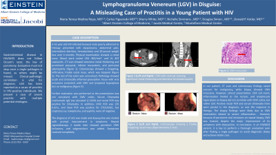
Case Description/Methods: A 31-year-old HIV-infected bisexual male, poorly adherent to therapy presented with dyspareunia, abdominal pain, intermittent diarrhea, hematochezia, and 20-pound weight loss over 4 months. Physical examination showed a rectal mass. Blood work noted CD4 365/mm3, and VL 467 copies/mL. CT scan showed extensive rectal thickening and prominent presacral, inferior mesenteric, and perirectal adenopathy (figures 1 and 2). A colonoscopy showed a fungating, infiltrative, friable rectal mass, which was biopsied (figure 3). The rest of the colon was uninvolved. Pathology showed acute and chronically inflamed granulation tissue with viral inclusions consistent with CMV and lymphoid hyperplasia but no malignancy (figure 4).
Further evaluation was performed as the presentation was not consistent with CMV colitis. Serum Chlamydia trachomatis IgG was elevated (1:1024) and rectal PCR was positive for Chlamydia. In addition, CMV PCR was 135 IU/mL, and Stool PCR was positive for ETEC/EAEC, but cryptococcus, toxoplasma, and syphilis, were all negative. The diagnosis of LGV was made and doxycycline was started with prompt improvement in symptoms. Repeat sigmoidoscopy 2 weeks later showed persistent CMV inclusions, and valganciclovir was added. Symptoms resolved completely.
Discussion: In our patient, CT scan and colonoscopy findings raised concern for malignancy, while biopsy showed CMV inclusions. However, clinical presentation, CD4 cell count, inflammation limited to the rectum, and lymphocytic hyperplasia on biopsy did not correlate with CMV colitis, but rather LGV. Positive rectal PCR and serum chlamydia titers were pivotal in the diagnosis, as was the response to therapy. The biopsy findings were likely due to CMV reactivation related to severe inflammation. However, because of persistent viral inclusions on repeat biopsy, CMV was treated, despite the major improvement of his symptoms with doxycycline. In conclusion, in an HIV/AIDS patient, it is key to perform a thorough evaluation even after finding a single pathogen to avoid diagnostic delays and achieve better care.

Figure: Figures 1 and 2: CTAP with contrast showing significant rectal thickening and extensive lymphadenopathy.
Figure 3: Colonoscopy showing a friable, fungating rectal mass (Approximately 5 cm).
Figure 4: CMV inclusions on biopsy.
Figure 3: Colonoscopy showing a friable, fungating rectal mass (Approximately 5 cm).
Figure 4: CMV inclusions on biopsy.
Disclosures:
Maria Teresa Medina Rojas indicated no relevant financial relationships.
Carlos Figueredo indicated no relevant financial relationships.
Douglas Simon indicated no relevant financial relationships.
Nichelle Simmons indicated no relevant financial relationships.
Sherrie White indicated no relevant financial relationships.
Donald Kotler: EMD Serono – Advisor or Review Panel Member.
Maria Teresa Medina Rojas, MD1, Carlos Figueredo, MD2, Douglas Simon, MD3, Nichelle Simmons, MD3, Sherrie White, MD3, Donald P. Kotler, MD4. P0388 - Lymphogranuloma Venereum (LGV) in Disguise: A Misleading Case of Proctitis in a Young Patient With HIV, ACG 2024 Annual Scientific Meeting Abstracts. Philadelphia, PA: American College of Gastroenterology.
