Sunday Poster Session
Category: Colon
P0389 - Rectal Xanthoma: A Case Report of a 56-Year-Old Female Patient
Sunday, October 27, 2024
3:30 PM - 7:00 PM ET
Location: Exhibit Hall E

Has Audio

Ismail Althunibat, MD
Saint Michael's Medical Center
Newark, NJ
Presenting Author(s)
Ismail Althunibat, MD1, Murad Qirem, MD2, Ahmad Alomari, MD3, Raed Atiyat, MD2, Yatinder Bains, MD2, Theodore DaCosta Jr, MD2, Gowthami Sai Kogilathota Jagirdhar, MD4, Muhammad Hussain, MD2
1Saint Michael's Medical Center, Newark, NJ; 2New York Medical College - Saint Michael's Medical Center, Newark, NJ; 3Henry Ford Hospital, Detroit, MI; 4Saint Michaels Medical Center, Newark, NJ
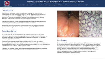
Introduction: Xanthomas are raised, waxy-looking, yellowish lesions resulting from accumulation of cholesterol and fats. Their most common location is the skin. However, though rare, they can occur in the gastrointestinal tract (GI). Typically, they are usually found incidentally on colonoscopic examinations, appearing as red polyps or occasionally as yellowish- white in formations with a size of 1-3 mm. They can be misdiagnosed for lipomas.
Although most commonly they are completely asymptomatic, some cases were reported with obstructive symptoms such as pain, abdominal distension and vomiting.
Histologically, rectal xanthomas consist of aggregates of foamy macrophages in the lamina propria, which are negative for mucin and cytokeratin but positive for CD68 immunostain.
Case Description/Methods: we present a case of a 55 year old gentleman with past medical history of diabetes type 2, hypertension, hyperlipidemia, and obesity, who presented to undergo screening colonoscopy. At the time of presentation, the patient exhibited no GI symptoms or complaints, and laboratory test results were within normal limits.
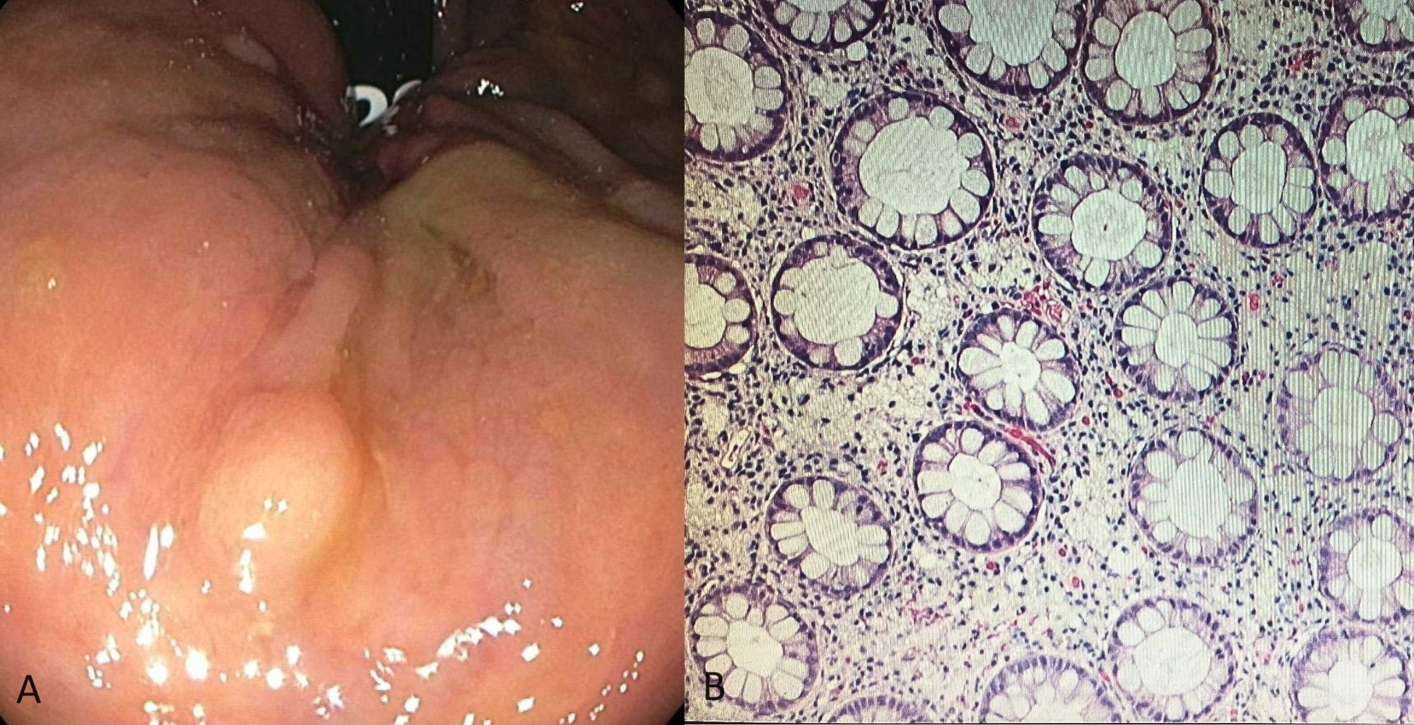
Colonoscopy was done and it showed diverticulosis throughout the colon, and multiple diminutive polyps, along with an 8 mm polyp that was found in the rectum. Biopsy was taken and histopathological evaluation of the 8mm polyp showed collections of foamy cells in the lamina propria suggestive of mucosal xanthoma, without dysplastic changes.
The patient was evaluated 2 weeks after the colonoscopy and was doing well with no complaints or symptoms, and will be followed based on the regular colonoscopy surveys.
Discussion: Some studies were focusing on the relationship between dyslipidaemia and the development of rectosigmoid xanthomas.one study highlighted the role of lipid metabolism abnormalities as the underlying cause of xanthomas with evidence suggesting that treating hyperlipidaemia can lead to the resolution of gastric xanthomas in some cases. Conversely, other studies found no significant association between gastric xanthomas and diabetes mellitus, hypercholesterolemia, or skin lesions. Further research is needed to explore the intricate relationship between xanthomas and lipid metabolism, shedding light on potential therapeutic avenues and management strategies.

Disclosures:
Ismail Althunibat, MD1, Murad Qirem, MD2, Ahmad Alomari, MD3, Raed Atiyat, MD2, Yatinder Bains, MD2, Theodore DaCosta Jr, MD2, Gowthami Sai Kogilathota Jagirdhar, MD4, Muhammad Hussain, MD2. P0389 - Rectal Xanthoma: A Case Report of a 56-Year-Old Female Patient, ACG 2024 Annual Scientific Meeting Abstracts. Philadelphia, PA: American College of Gastroenterology.
1Saint Michael's Medical Center, Newark, NJ; 2New York Medical College - Saint Michael's Medical Center, Newark, NJ; 3Henry Ford Hospital, Detroit, MI; 4Saint Michaels Medical Center, Newark, NJ
Introduction: Xanthomas are raised, waxy-looking, yellowish lesions resulting from accumulation of cholesterol and fats. Their most common location is the skin. However, though rare, they can occur in the gastrointestinal tract (GI). Typically, they are usually found incidentally on colonoscopic examinations, appearing as red polyps or occasionally as yellowish- white in formations with a size of 1-3 mm. They can be misdiagnosed for lipomas.
Although most commonly they are completely asymptomatic, some cases were reported with obstructive symptoms such as pain, abdominal distension and vomiting.
Histologically, rectal xanthomas consist of aggregates of foamy macrophages in the lamina propria, which are negative for mucin and cytokeratin but positive for CD68 immunostain.
Case Description/Methods: we present a case of a 55 year old gentleman with past medical history of diabetes type 2, hypertension, hyperlipidemia, and obesity, who presented to undergo screening colonoscopy. At the time of presentation, the patient exhibited no GI symptoms or complaints, and laboratory test results were within normal limits.
Colonoscopy was done and it showed diverticulosis throughout the colon, and multiple diminutive polyps, along with an 8 mm polyp that was found in the rectum. Biopsy was taken and histopathological evaluation of the 8mm polyp showed collections of foamy cells in the lamina propria suggestive of mucosal xanthoma, without dysplastic changes.
The patient was evaluated 2 weeks after the colonoscopy and was doing well with no complaints or symptoms, and will be followed based on the regular colonoscopy surveys.
Discussion: Some studies were focusing on the relationship between dyslipidaemia and the development of rectosigmoid xanthomas.one study highlighted the role of lipid metabolism abnormalities as the underlying cause of xanthomas with evidence suggesting that treating hyperlipidaemia can lead to the resolution of gastric xanthomas in some cases. Conversely, other studies found no significant association between gastric xanthomas and diabetes mellitus, hypercholesterolemia, or skin lesions. Further research is needed to explore the intricate relationship between xanthomas and lipid metabolism, shedding light on potential therapeutic avenues and management strategies.

Figure: An 8 mm yellow-whitish polyp found at the rectum (A). Microscopic evaluation showed large vaculated macrophages filled with lipid droplets (B).
Disclosures:
Ismail Althunibat indicated no relevant financial relationships.
Murad Qirem indicated no relevant financial relationships.
Ahmad Alomari indicated no relevant financial relationships.
Raed Atiyat indicated no relevant financial relationships.
Yatinder Bains indicated no relevant financial relationships.
Theodore DaCosta Jr indicated no relevant financial relationships.
Gowthami Sai Kogilathota Jagirdhar indicated no relevant financial relationships.
Muhammad Hussain indicated no relevant financial relationships.
Ismail Althunibat, MD1, Murad Qirem, MD2, Ahmad Alomari, MD3, Raed Atiyat, MD2, Yatinder Bains, MD2, Theodore DaCosta Jr, MD2, Gowthami Sai Kogilathota Jagirdhar, MD4, Muhammad Hussain, MD2. P0389 - Rectal Xanthoma: A Case Report of a 56-Year-Old Female Patient, ACG 2024 Annual Scientific Meeting Abstracts. Philadelphia, PA: American College of Gastroenterology.
