Sunday Poster Session
Category: Colon
P0306 - A Case of Foley Catheter Protrusion Through a Colovesical Fistula
Sunday, October 27, 2024
3:30 PM - 7:00 PM ET
Location: Exhibit Hall E

Has Audio

Ryan Plunkett, MD
Saint Louis University School of Medicine
St. Louis, MO
Presenting Author(s)
Ryan Plunkett, MD1, Hayden Rotramel, MD2, Allison Shildt, BS1, Christopher Nguyen, DO1, Zain Raza, MD1, Michelle Baliss, DO2, Jered Schenk, MD3, Manar Shmais, MD4
1Saint Louis University School of Medicine, St. Louis, MO; 2SSM Health Saint Louis University Hospital, St. Louis, MO; 3Medical University of South Carolina, Charleston, SC; 4American University of Beirut, Beirut, Beyrouth, Lebanon
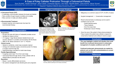
Introduction: A colovesical fistula (CVF) is an abnormal communication between the bowel and the bladder. Here, we describe a case of CVF due to malignancy and highlight the need for further advancement in management options for this disorder.
Case Description/Methods: A 65 year-old male with a history of metastatic prostate cancer was admitted for failure to thrive and altered mental status. Imaging showed a large fistula between the walls of the bladder and the rectum and a necrotic collection in the prostate bed communicating with the CVF. Labs were notable for WBC=21.6k, Escherichia coli and Citrobacter freundii bacteremia, and an undetectable prostate specific antigen. Antibiotics and evaluation of the rectal mass were initiated. Urology and colorectal surgery were concerned that pursuit of a transurethral biopsy or aggressive surgical management such as a diverting ostomy would be high risk and gastroenterology was consulted for rectal mass biopsy.
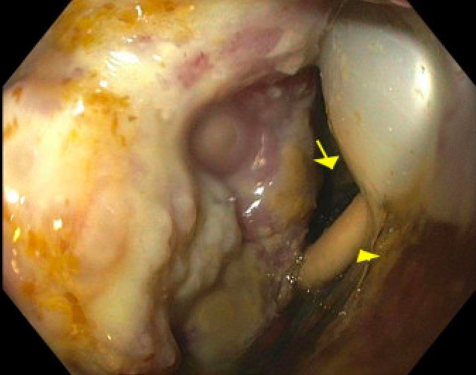
Physical exam demonstrated a cachectic patient with straw-colored rectal output. Flexible sigmoidoscopy exhibited a fungating rectal mass 10 cm from the anal verge, with a colovesical fistula, through which a Foley catheter with its inflated balloon protruded into the lumen. The rectal output was determined to be urine given this communication with the bladder. Rectal mass biopsies confirmed poorly differentiated carcinoma. No curative management was attempted for the fistula or the malignancy given the overall prognosis. The patient ultimately declined palliative care. Percutaneous nephrostomy tubes were placed and he was discharged to a skilled nursing facility.
Discussion: As demonstrated by this case, malignancy is a common cause of CVF (10-20% of cases). Conservative management with antibiotics and corticosteroids can be trialed, but surgical intervention is ultimately the preferred treatment. However, recent advancements in endoscopic technology (such as gastrointestinal stents, tissue sealants, over the scope clips, and endoscopic vacuum therapy) have made endoscopic management of CVFs a possibility.
Given the size of our patient’s fistula (as evidenced by the Foley catheter extending through the fistula and into the colonic lumen), he was unlikely to have responded to conservative management, yet was also determined to be a poor surgical candidate. This case highlights the need for continued advancements in endoscopic management of this condition as it offers a safer option for patients with contraindications to the current standards of treatment.

Disclosures:
Ryan Plunkett, MD1, Hayden Rotramel, MD2, Allison Shildt, BS1, Christopher Nguyen, DO1, Zain Raza, MD1, Michelle Baliss, DO2, Jered Schenk, MD3, Manar Shmais, MD4. P0306 - A Case of Foley Catheter Protrusion Through a Colovesical Fistula, ACG 2024 Annual Scientific Meeting Abstracts. Philadelphia, PA: American College of Gastroenterology.
1Saint Louis University School of Medicine, St. Louis, MO; 2SSM Health Saint Louis University Hospital, St. Louis, MO; 3Medical University of South Carolina, Charleston, SC; 4American University of Beirut, Beirut, Beyrouth, Lebanon
Introduction: A colovesical fistula (CVF) is an abnormal communication between the bowel and the bladder. Here, we describe a case of CVF due to malignancy and highlight the need for further advancement in management options for this disorder.
Case Description/Methods: A 65 year-old male with a history of metastatic prostate cancer was admitted for failure to thrive and altered mental status. Imaging showed a large fistula between the walls of the bladder and the rectum and a necrotic collection in the prostate bed communicating with the CVF. Labs were notable for WBC=21.6k, Escherichia coli and Citrobacter freundii bacteremia, and an undetectable prostate specific antigen. Antibiotics and evaluation of the rectal mass were initiated. Urology and colorectal surgery were concerned that pursuit of a transurethral biopsy or aggressive surgical management such as a diverting ostomy would be high risk and gastroenterology was consulted for rectal mass biopsy.
Physical exam demonstrated a cachectic patient with straw-colored rectal output. Flexible sigmoidoscopy exhibited a fungating rectal mass 10 cm from the anal verge, with a colovesical fistula, through which a Foley catheter with its inflated balloon protruded into the lumen. The rectal output was determined to be urine given this communication with the bladder. Rectal mass biopsies confirmed poorly differentiated carcinoma. No curative management was attempted for the fistula or the malignancy given the overall prognosis. The patient ultimately declined palliative care. Percutaneous nephrostomy tubes were placed and he was discharged to a skilled nursing facility.
Discussion: As demonstrated by this case, malignancy is a common cause of CVF (10-20% of cases). Conservative management with antibiotics and corticosteroids can be trialed, but surgical intervention is ultimately the preferred treatment. However, recent advancements in endoscopic technology (such as gastrointestinal stents, tissue sealants, over the scope clips, and endoscopic vacuum therapy) have made endoscopic management of CVFs a possibility.
Given the size of our patient’s fistula (as evidenced by the Foley catheter extending through the fistula and into the colonic lumen), he was unlikely to have responded to conservative management, yet was also determined to be a poor surgical candidate. This case highlights the need for continued advancements in endoscopic management of this condition as it offers a safer option for patients with contraindications to the current standards of treatment.

Figure: Figure 1. The colovesical fistula (arrow) and the Foley catheter extending from the fistula (arrowhead).
Disclosures:
Ryan Plunkett indicated no relevant financial relationships.
Hayden Rotramel indicated no relevant financial relationships.
Allison Shildt indicated no relevant financial relationships.
Christopher Nguyen indicated no relevant financial relationships.
Zain Raza indicated no relevant financial relationships.
Michelle Baliss indicated no relevant financial relationships.
Jered Schenk indicated no relevant financial relationships.
Manar Shmais indicated no relevant financial relationships.
Ryan Plunkett, MD1, Hayden Rotramel, MD2, Allison Shildt, BS1, Christopher Nguyen, DO1, Zain Raza, MD1, Michelle Baliss, DO2, Jered Schenk, MD3, Manar Shmais, MD4. P0306 - A Case of Foley Catheter Protrusion Through a Colovesical Fistula, ACG 2024 Annual Scientific Meeting Abstracts. Philadelphia, PA: American College of Gastroenterology.
