Sunday Poster Session
Category: Biliary/Pancreas
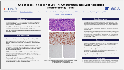
P0155 - One of These Things Is Not Like the Other: Primary Bile Duct-Associated Neuroendocrine Tumor
Sunday, October 27, 2024
3:30 PM - 7:00 PM ET
Location: Exhibit Hall E

Has Audio
- BP
Bishal Paudel, MD
University of Florida
Gainesville, FL
Presenting Author(s)
Bishal Paudel, MD1, Andrew Kleehammer, DO2, Jennifer Fieber, MD2, Ibrahim Nassour, MD1, Hassam Cheema, MD1, Aleksey Novikov, MD2
1University of Florida, Gainesville, FL; 2University of Florida College of Medicine, Gainesville, FL
Introduction: Neuroendocrine tumor (NET) is a rare neoplasm that most commonly occurs in the gastrointestinal tract. These tumors arise from enterochromaffin cells and can be functional or non-functional. Primary NET of the bile duct is very uncommon with only a few cases reported in the literature and account for less than 0.5% of all primary NETs. Biliary tree NETs generally have good prognosis, and complete surgical excision offers optimal treatment.
Case Description/Methods: A 38-year-old female with past medical history of poorly controlled type II diabetes, metabolic dysfunction associated steatotic liver disease, depression, GERD, intermittent gastroparesis and hypertriglyceridemia presented to the emergency department with complaints of hyperglycemia and pruritis. Initial labs were significant for AST 113 IU/L, ALT 200 IU/L, total bilirubin 2.4 mg/dL, alkaline phosphatase 725 IU/L, triglyceride 593 mg/dL and blood glucose 324 mg/dL. CT Abdomen Pelvis showed intrahepatic biliary ductal dilation with no obstructing lesion identified. MRCP upon discharge showed a mass centered in the porta hepatis region causing compression of the common bile duct (CBD).
EUS showed a mass arising from CBD. There was no evidence of pancreatic or duodenal involvement. ERCP with cholangioscopy showed a biliary stricture in the common bile duct which was biopsied. Pathology from both EUS and ERCP showed well-differentiated NET. Further workup showed elevated chromogranin A and only mildly elevated gastrin and glucagon levels. Vasoactive intestinal peptide and urine 5-hydroxyindoleacetic acid levels were normal. PET CT Dotatate scan showed intense uptake in the CBD, without involvement of the pancreas. The patient subsequently underwent periportal mass resection, extrahepatic bile duct resection with Roux-en-y hepaticojejunostomy and reconstruction of the right hepatic artery. The pathology of the mass showed a well-differentiated NET with positive chromogranin A and negative gastrin and glucagon staining.
Discussion: Well differentiated primary NET of the bile duct is a rare neoplasm that carries a good prognosis. It was clinically challenging to distinguish between a primary bile duct associated NET versus a porta hepatis lymph node invaded by metastatic NET. In this case, well differentiated pathology and reassuring results of EUS and PET-CT led to a curative, less invasive surgery. Well differentiated primary NET of the bile duct should be considered in patients with masses in the porta hepatis region.

Disclosures:
Bishal Paudel, MD1, Andrew Kleehammer, DO2, Jennifer Fieber, MD2, Ibrahim Nassour, MD1, Hassam Cheema, MD1, Aleksey Novikov, MD2. P0155 - One of These Things Is Not Like the Other: Primary Bile Duct-Associated Neuroendocrine Tumor, ACG 2024 Annual Scientific Meeting Abstracts. Philadelphia, PA: American College of Gastroenterology.
1University of Florida, Gainesville, FL; 2University of Florida College of Medicine, Gainesville, FL
Introduction: Neuroendocrine tumor (NET) is a rare neoplasm that most commonly occurs in the gastrointestinal tract. These tumors arise from enterochromaffin cells and can be functional or non-functional. Primary NET of the bile duct is very uncommon with only a few cases reported in the literature and account for less than 0.5% of all primary NETs. Biliary tree NETs generally have good prognosis, and complete surgical excision offers optimal treatment.
Case Description/Methods: A 38-year-old female with past medical history of poorly controlled type II diabetes, metabolic dysfunction associated steatotic liver disease, depression, GERD, intermittent gastroparesis and hypertriglyceridemia presented to the emergency department with complaints of hyperglycemia and pruritis. Initial labs were significant for AST 113 IU/L, ALT 200 IU/L, total bilirubin 2.4 mg/dL, alkaline phosphatase 725 IU/L, triglyceride 593 mg/dL and blood glucose 324 mg/dL. CT Abdomen Pelvis showed intrahepatic biliary ductal dilation with no obstructing lesion identified. MRCP upon discharge showed a mass centered in the porta hepatis region causing compression of the common bile duct (CBD).
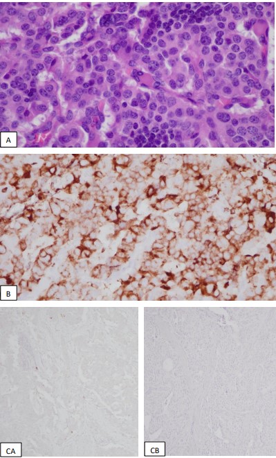
EUS showed a mass arising from CBD. There was no evidence of pancreatic or duodenal involvement. ERCP with cholangioscopy showed a biliary stricture in the common bile duct which was biopsied. Pathology from both EUS and ERCP showed well-differentiated NET. Further workup showed elevated chromogranin A and only mildly elevated gastrin and glucagon levels. Vasoactive intestinal peptide and urine 5-hydroxyindoleacetic acid levels were normal. PET CT Dotatate scan showed intense uptake in the CBD, without involvement of the pancreas. The patient subsequently underwent periportal mass resection, extrahepatic bile duct resection with Roux-en-y hepaticojejunostomy and reconstruction of the right hepatic artery. The pathology of the mass showed a well-differentiated NET with positive chromogranin A and negative gastrin and glucagon staining.
Discussion: Well differentiated primary NET of the bile duct is a rare neoplasm that carries a good prognosis. It was clinically challenging to distinguish between a primary bile duct associated NET versus a porta hepatis lymph node invaded by metastatic NET. In this case, well differentiated pathology and reassuring results of EUS and PET-CT led to a curative, less invasive surgery. Well differentiated primary NET of the bile duct should be considered in patients with masses in the porta hepatis region.

Figure: Fig 1: A- Well differentiated neuroendocrine tumor with classic “salt and pepper” chromatin
B- Neuroendocrine tumor with chromogranin stain
CA- Negative gastrin stain CB- Negative glucagon strain
B- Neuroendocrine tumor with chromogranin stain
CA- Negative gastrin stain CB- Negative glucagon strain
Disclosures:
Bishal Paudel indicated no relevant financial relationships.
Andrew Kleehammer indicated no relevant financial relationships.
Jennifer Fieber indicated no relevant financial relationships.
Ibrahim Nassour indicated no relevant financial relationships.
Hassam Cheema indicated no relevant financial relationships.
Aleksey Novikov indicated no relevant financial relationships.
Bishal Paudel, MD1, Andrew Kleehammer, DO2, Jennifer Fieber, MD2, Ibrahim Nassour, MD1, Hassam Cheema, MD1, Aleksey Novikov, MD2. P0155 - One of These Things Is Not Like the Other: Primary Bile Duct-Associated Neuroendocrine Tumor, ACG 2024 Annual Scientific Meeting Abstracts. Philadelphia, PA: American College of Gastroenterology.
