Sunday Poster Session
Category: Biliary/Pancreas
P0168 - Biopsy Proven Sarcoidosis Presenting as Secondary Sclerosing Cholangitis
Sunday, October 27, 2024
3:30 PM - 7:00 PM ET
Location: Exhibit Hall E

Has Audio
- RH
Rwan Hayek
Medical College of Georgia at Augusta University
Augusta, GA
Presenting Author(s)
Rwan Hayek, 1, Angela Barnes, MD, MBChB1, Tanya Ratnani, MBBS2, Subbaramiah Sridhar, MBBS, MPH3, Abdul-Rahman Abualruz, MD2
1Medical College of Georgia at Augusta University, Augusta, GA; 2Medical College of Georgia, Augusta University, Augusta, GA; 3Augusta University, Augusta, GA
Introduction: Sarcoidosis is an inflammatory disease which affects multiple organs, the lungs being the most commonly involved organs. Bile duct involvement is rare. We present a case of biopsy proven sarcoidosis masquerading as Secondary Sclerosing Cholangitis.
Case Description/Methods: A 57-year-old female with a history of cholecystectomy was referred to the GI clinic for evaluation of a 6 month’s history of epigastric pain, weight loss, and elevated alkaline phosphatase of 802 IU/L and CA 19-9 level of 78 U/mL.
An upper endoscopy revealed an extrinsic pulsating bulge compressing the duodenal bulb.
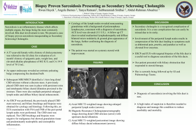
Subsequent MRI/MRCP identified a 1.6cm long distal CBD stricture without a discrete mass. A previously placed CBD stent was seen in place with persistent extra and intrahepatic biliary ductal dilatation proximal to the stricture. There were also multiple periportal enlarged lymph nodes and diffuse biliary ductal enhancement. An ERCP was performed, the previously placed CBD stent removed and the biliary brushings and biopsies were obtained for cytology and histology. Following this, an EUS examination with FNA and FNB of the peri-portal lymph nodes were performed. The CBD stent was replaced. The CBD brushings and biopsies were negative for malignancy but showed granulation tissue and predominantly neutrophilic and eosinophilic inflammation. Cytology of the lymph nodes revealed noncaseating granulomas with a background of polymorphous lymphocytes, consistent with a diagnosis of sarcoidosis. ACE level was elevated (111 U/L) . A follow up CT chest revealed mediastinal lymphadenopathy and diffuse bilateral micro nodularity & ground glass appearance of the lungs, further confirming the diagnosis of sarcoidosis. The patient was started on systemic steroid with improvement, and she is currently being followed up by GI and Pulmonology Teams.
Discussion: Secondary cholangitis is a recognized complication of sarcoidosis. It is a rare complication that can easily be missed due to its rarity. Involvement of the periportal lymph nodes results in compression of the bile duct leading to symptoms such as abdominal pain, pruritis and jaundice as well as elevated liver enzymes. ERCP and EUS with targeted biopsies of the bile duct is a useful tool to diagnose sarcoidosis of the bile ducts. Our patient presented with biliary obstruction that responded to steroid therapy. A high index of suspicion is therefore needed to diagnose and manage this condition to reduce morbidity and mortality.

Disclosures:
Rwan Hayek, 1, Angela Barnes, MD, MBChB1, Tanya Ratnani, MBBS2, Subbaramiah Sridhar, MBBS, MPH3, Abdul-Rahman Abualruz, MD2. P0168 - Biopsy Proven Sarcoidosis Presenting as Secondary Sclerosing Cholangitis, ACG 2024 Annual Scientific Meeting Abstracts. Philadelphia, PA: American College of Gastroenterology.
1Medical College of Georgia at Augusta University, Augusta, GA; 2Medical College of Georgia, Augusta University, Augusta, GA; 3Augusta University, Augusta, GA
Introduction: Sarcoidosis is an inflammatory disease which affects multiple organs, the lungs being the most commonly involved organs. Bile duct involvement is rare. We present a case of biopsy proven sarcoidosis masquerading as Secondary Sclerosing Cholangitis.
Case Description/Methods: A 57-year-old female with a history of cholecystectomy was referred to the GI clinic for evaluation of a 6 month’s history of epigastric pain, weight loss, and elevated alkaline phosphatase of 802 IU/L and CA 19-9 level of 78 U/mL.
An upper endoscopy revealed an extrinsic pulsating bulge compressing the duodenal bulb.
Subsequent MRI/MRCP identified a 1.6cm long distal CBD stricture without a discrete mass. A previously placed CBD stent was seen in place with persistent extra and intrahepatic biliary ductal dilatation proximal to the stricture. There were also multiple periportal enlarged lymph nodes and diffuse biliary ductal enhancement. An ERCP was performed, the previously placed CBD stent removed and the biliary brushings and biopsies were obtained for cytology and histology. Following this, an EUS examination with FNA and FNB of the peri-portal lymph nodes were performed. The CBD stent was replaced. The CBD brushings and biopsies were negative for malignancy but showed granulation tissue and predominantly neutrophilic and eosinophilic inflammation. Cytology of the lymph nodes revealed noncaseating granulomas with a background of polymorphous lymphocytes, consistent with a diagnosis of sarcoidosis. ACE level was elevated (111 U/L) . A follow up CT chest revealed mediastinal lymphadenopathy and diffuse bilateral micro nodularity & ground glass appearance of the lungs, further confirming the diagnosis of sarcoidosis. The patient was started on systemic steroid with improvement, and she is currently being followed up by GI and Pulmonology Teams.
Discussion: Secondary cholangitis is a recognized complication of sarcoidosis. It is a rare complication that can easily be missed due to its rarity. Involvement of the periportal lymph nodes results in compression of the bile duct leading to symptoms such as abdominal pain, pruritis and jaundice as well as elevated liver enzymes. ERCP and EUS with targeted biopsies of the bile duct is a useful tool to diagnose sarcoidosis of the bile ducts. Our patient presented with biliary obstruction that responded to steroid therapy. A high index of suspicion is therefore needed to diagnose and manage this condition to reduce morbidity and mortality.

Figure: A) Axial MRI T2 weighted image showing enlarged periportal lymph nodes (arrows).
B) Magnetic Resonance Cholangiopancreatography image showing distal CBD stricture (arrow) with upstream ductal dilatation.
C) Axial MRI T1 weighted postcontrast image showing diffuse biliary ductal enhancement (arrows).
B) Magnetic Resonance Cholangiopancreatography image showing distal CBD stricture (arrow) with upstream ductal dilatation.
C) Axial MRI T1 weighted postcontrast image showing diffuse biliary ductal enhancement (arrows).
Disclosures:
Rwan Hayek indicated no relevant financial relationships.
Angela Barnes indicated no relevant financial relationships.
Tanya Ratnani indicated no relevant financial relationships.
Subbaramiah Sridhar indicated no relevant financial relationships.
Abdul-Rahman Abualruz indicated no relevant financial relationships.
Rwan Hayek, 1, Angela Barnes, MD, MBChB1, Tanya Ratnani, MBBS2, Subbaramiah Sridhar, MBBS, MPH3, Abdul-Rahman Abualruz, MD2. P0168 - Biopsy Proven Sarcoidosis Presenting as Secondary Sclerosing Cholangitis, ACG 2024 Annual Scientific Meeting Abstracts. Philadelphia, PA: American College of Gastroenterology.
