Sunday Poster Session
Category: Biliary/Pancreas
P0082 - Delayed Presentation of Bile Leak Following Laparoscopic Cholecystectomy: A Case Report
Sunday, October 27, 2024
3:30 PM - 7:00 PM ET
Location: Exhibit Hall E

Has Audio
- IB
Imane Bouhali
Weill Cornell Medicine
New York, NY
Presenting Author(s)
Imane Bouhali, 1, Kyle Liang, PA1, Mark Hanscom, MD1, Enad Dawod, MD1, Anam Rizvi, MD1, David Wan, MD2, SriHari Mahadev, MD2
1Weill Cornell Medicine, New York, NY; 2New York-Presbyterian / Weill Cornell Medical Center, New York, NY
Introduction: Laparoscopic cholecystectomy (CCY) is the gold-standard surgical intervention for management of a variety of gallbladder pathologies. While generally safe, complications include bile leaks that can progress to conditions such as bilomas, abscesses, and sepsis if left untreated. Recognizing and managing these leaks early is crucial in determining patient outcome.
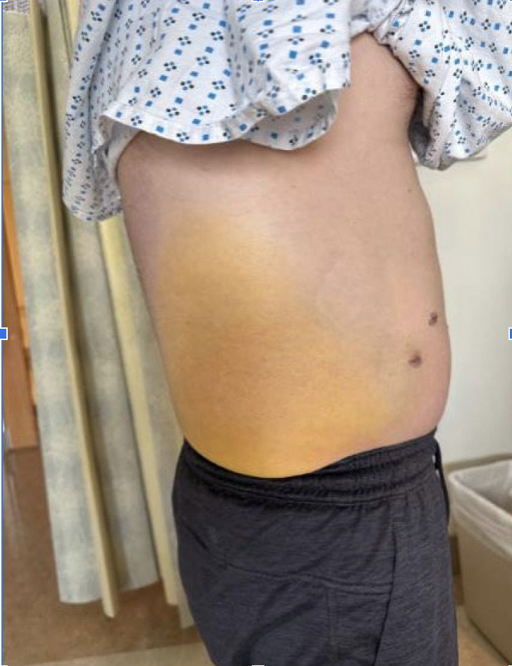
Case Description/Methods: A 46 year old male with a history of GERD and gallbladder hyperkinesia was readmitted one day post-elective CCY for abdominal pain. On exam, patient’s right lower quadrant was tender to palpation without rebound or guarding. Labs were notable for WBC 26.32x10(3)uL , Tbili 2.1 (direct 0.9 mg/dL), AST 27, ALT 55, ALP 66 U/L. Lipase 26 U/L. CT w/ IV contrast showed postsurgical changes including fat stranding within gallbladder fossa and hyperenhancement of the common bile duct. Subsequent magnetic resonance cholangiopancreatography (MRCP) revealed perihepatic and trace amounts of fluid in gallbladder fossa. Hepatobiliary iminodiacetic acid (HIDA) scan was negative for bile leak. However, given persistent symptoms, decision was made to proceed with endoscopic ultrasound (EUS) which showed bilious ascites in gallbladder fossa. Subsequent Endoscopic retrograde cholangiopancreatography (ERCP) revealed a bile leak originating from the remnant cystic duct which was managed with biliary sphincterotomy and placement of 10 Fr x 5 cm straight plastic stent. Patient reported resolving abdominal pain but noted new icterus marginatus localized to the right flank. Patient was discharged but readmitted 2 weeks later with 2 days of epigastric pain. CT showed a new 6.6 cm thick walled fluid collection in the gallbladder fossa, consistent with a contained leak from the cystic duct stump. Patient underwent EUS/ERCP showcasing persistent leak. The stent was exchanged to an 8 mm x 6 cm covered metal stent and collection was drained into the duodenum with a 10 x 10 mm lumen-apposing metal stent. Subsequent CT showed a resolution of the collection.
Discussion: Bile leaks are rare complications of CCY. Diagnosis of a bile leak requires strong clinical suspicion, such as persistent abdominal pain and rarely icterus marginatum. HIDA scans can evaluate these leaks, but can be deceptively normal. As such, if clinical suspicion is high, various modalities should be used to assess pathology as none is perfectly sensitive. Endoscopic stenting is generally an effective treatment, with tailoring of treatment to size and location of the leak.

Disclosures:
Imane Bouhali, 1, Kyle Liang, PA1, Mark Hanscom, MD1, Enad Dawod, MD1, Anam Rizvi, MD1, David Wan, MD2, SriHari Mahadev, MD2. P0082 - Delayed Presentation of Bile Leak Following Laparoscopic Cholecystectomy: A Case Report, ACG 2024 Annual Scientific Meeting Abstracts. Philadelphia, PA: American College of Gastroenterology.
1Weill Cornell Medicine, New York, NY; 2New York-Presbyterian / Weill Cornell Medical Center, New York, NY
Introduction: Laparoscopic cholecystectomy (CCY) is the gold-standard surgical intervention for management of a variety of gallbladder pathologies. While generally safe, complications include bile leaks that can progress to conditions such as bilomas, abscesses, and sepsis if left untreated. Recognizing and managing these leaks early is crucial in determining patient outcome.
Case Description/Methods: A 46 year old male with a history of GERD and gallbladder hyperkinesia was readmitted one day post-elective CCY for abdominal pain. On exam, patient’s right lower quadrant was tender to palpation without rebound or guarding. Labs were notable for WBC 26.32x10(3)uL , Tbili 2.1 (direct 0.9 mg/dL), AST 27, ALT 55, ALP 66 U/L. Lipase 26 U/L. CT w/ IV contrast showed postsurgical changes including fat stranding within gallbladder fossa and hyperenhancement of the common bile duct. Subsequent magnetic resonance cholangiopancreatography (MRCP) revealed perihepatic and trace amounts of fluid in gallbladder fossa. Hepatobiliary iminodiacetic acid (HIDA) scan was negative for bile leak. However, given persistent symptoms, decision was made to proceed with endoscopic ultrasound (EUS) which showed bilious ascites in gallbladder fossa. Subsequent Endoscopic retrograde cholangiopancreatography (ERCP) revealed a bile leak originating from the remnant cystic duct which was managed with biliary sphincterotomy and placement of 10 Fr x 5 cm straight plastic stent. Patient reported resolving abdominal pain but noted new icterus marginatus localized to the right flank. Patient was discharged but readmitted 2 weeks later with 2 days of epigastric pain. CT showed a new 6.6 cm thick walled fluid collection in the gallbladder fossa, consistent with a contained leak from the cystic duct stump. Patient underwent EUS/ERCP showcasing persistent leak. The stent was exchanged to an 8 mm x 6 cm covered metal stent and collection was drained into the duodenum with a 10 x 10 mm lumen-apposing metal stent. Subsequent CT showed a resolution of the collection.
Discussion: Bile leaks are rare complications of CCY. Diagnosis of a bile leak requires strong clinical suspicion, such as persistent abdominal pain and rarely icterus marginatum. HIDA scans can evaluate these leaks, but can be deceptively normal. As such, if clinical suspicion is high, various modalities should be used to assess pathology as none is perfectly sensitive. Endoscopic stenting is generally an effective treatment, with tailoring of treatment to size and location of the leak.

Figure: Icterus marginatum as a result of bile leak post-CCY
Disclosures:
Imane Bouhali indicated no relevant financial relationships.
Kyle Liang indicated no relevant financial relationships.
Mark Hanscom indicated no relevant financial relationships.
Enad Dawod indicated no relevant financial relationships.
Anam Rizvi indicated no relevant financial relationships.
David Wan indicated no relevant financial relationships.
SriHari Mahadev: Boston Scientific – Consultant. Conmed – Consultant.
Imane Bouhali, 1, Kyle Liang, PA1, Mark Hanscom, MD1, Enad Dawod, MD1, Anam Rizvi, MD1, David Wan, MD2, SriHari Mahadev, MD2. P0082 - Delayed Presentation of Bile Leak Following Laparoscopic Cholecystectomy: A Case Report, ACG 2024 Annual Scientific Meeting Abstracts. Philadelphia, PA: American College of Gastroenterology.
